1GMC | pdb_00001gmc

THE X-RAY CRYSTAL STRUCTURE OF THE TETRAHEDRAL INTERMEDIATE OF GAMMA-CHYMOTRYPSIN IN HEXANE


Experimental Data Snapshot

  • Method: X-RAY DIFFRACTION
  • Resolution: 2.20 Å
  • R-Value Work: 
    0.176 (Depositor) 
  • R-Value Observed: 
    0.176 (Depositor) 

wwPDB Validation 3D Report Full Report

Validation slider image for 1GMC

This is version 1.6 of the entry. See complete history

Literature

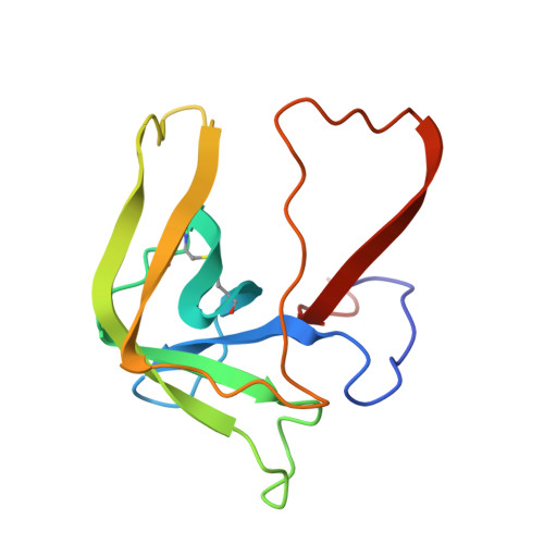
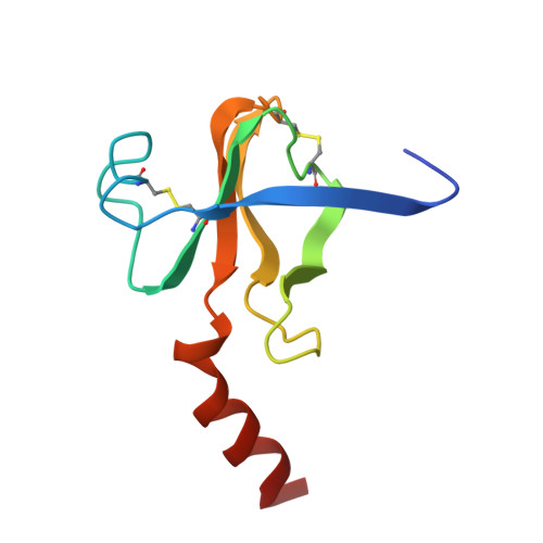
X-ray crystal structure of gamma-chymotrypsin in hexane.

Yennawar, N.H.Yennawar, H.P.Farber, G.K.

(1994) Biochemistry 33: 7326-7336

  • DOI: https://doi.org/10.1021/bi00189a038
  • Primary Citation Related Structures: 
    1GMC, 1GMD

  • PubMed Abstract: 

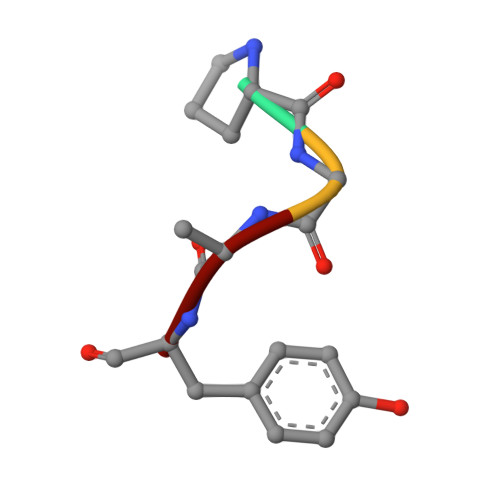
    Crystals of gamma-chymotrypsin grown in aqueous solution were soaked in n-hexane, and the structures of both the soaked and the native crystals were determined to 2.2-A resolution. Seven hexane molecules and 130 water molecules were found in the hexane-soaked crystals. Two of the seven hexane molecules are found near the active site, and the rest are close to hydrophobic regions on or near the surface of the enzyme. In the hexane structure, water molecules that were not observed in the native structure form a clathrate around one of the hexane molecules. Only 97 water molecules were found in the native structure. The temperature factors for atoms in the hexane environment are lower than those in the aqueous environment. There are significant changes between the two structures in the side chains of both polar and neutral residues, particularly in the vicinity of the hexane molecules. These changes have perturbed the hydrogen-bonding patterns. The electron density for the peptide bound in the active site has been dramatically altered in hexane and appears to be tetrahedral at the carbon that is covalently bound to Ser 195. The crystalline enzyme retains its active conformation in the nonpolar medium and can catalyze both hydrolysis and synthesis reactions in hexane.


  • Organizational Affiliation
    • Department of Chemistry, Pennsylvania State University, University Park 16802.

Macromolecule Content 

  • Total Structure Weight: 25.67 kDa 
  • Atom Count: 1,863 
  • Modeled Residue Count: 241 
  • Deposited Residue Count: 245 
  • Unique protein chains: 4

Macromolecules

Find similar proteins by:  Sequence   |   3D Structure  
Entity ID: 1
MoleculeChains  Sequence LengthOrganismDetailsImage
GAMMA-CHYMOTRYPSIN AA [auth E]13Bos taurusMutation(s): 0 
EC: 3.4.21.1
UniProt
Find proteins for P00766 (Bos taurus)
Explore P00766 
Go to UniProtKB:  P00766
Entity Groups
Sequence Clusters30% Identity50% Identity70% Identity90% Identity95% Identity100% Identity
UniProt GroupP00766
Sequence Annotations
Expand
Reference Sequence
Find similar proteins by:|  3D Structure
Entity ID: 2
MoleculeChains  Sequence LengthOrganismDetailsImage
GAMMA-CHYMOTRYPSIN AB [auth F]131Bos taurusMutation(s): 0 
EC: 3.4.21.1
UniProt
Find proteins for P00766 (Bos taurus)
Explore P00766 
Go to UniProtKB:  P00766
Entity Groups
Sequence Clusters30% Identity50% Identity70% Identity90% Identity95% Identity100% Identity
UniProt GroupP00766
Sequence Annotations
Expand
Reference Sequence
Find similar proteins by:|  3D Structure
Entity ID: 3
MoleculeChains  Sequence LengthOrganismDetailsImage
GAMMA-CHYMOTRYPSIN AC [auth G]97Bos taurusMutation(s): 0 
EC: 3.4.21.1
UniProt
Find proteins for P00766 (Bos taurus)
Explore P00766 
Go to UniProtKB:  P00766
Entity Groups
Sequence Clusters30% Identity50% Identity70% Identity90% Identity95% Identity100% Identity
UniProt GroupP00766
Sequence Annotations
Expand
Reference Sequence
Find similar proteins by:  Sequence   |   3D Structure  
Entity ID: 4
MoleculeChains  Sequence LengthOrganismDetailsImage
PRO GLY ALA TYR PEPTIDED [auth B]4N/AMutation(s): 0 

Experimental Data & Validation

Experimental Data

  • Method: X-RAY DIFFRACTION
  • Resolution: 2.20 Å
  • R-Value Work:  0.176 (Depositor) 
  • R-Value Observed: 0.176 (Depositor) 
Space Group: P 42 21 2
Unit Cell:
Length ( Å )Angle ( ˚ )
a = 69.54α = 90
b = 69.52β = 90
c = 98.01γ = 90
Software Package:
Software NamePurpose
X-PLORrefinement

Structure Validation

View Full Validation Report


Entry History 

Deposition Data

Revision History  (Full details and data files)

  • Version 1.0: 1993-10-31
    Type: Initial release
  • Version 1.1: 2008-03-24
    Changes: Version format compliance
  • Version 1.2: 2011-07-13
    Changes: Atomic model, Database references, Derived calculations, Non-polymer description, Structure summary, Version format compliance
  • Version 1.3: 2012-12-12
    Changes: Other
  • Version 1.4: 2013-03-13
    Changes: Other
  • Version 1.5: 2024-06-05
    Changes: Data collection, Database references, Derived calculations, Other
  • Version 1.6: 2024-11-20
    Changes: Structure summary