Contribution of polar groups in the interior of a protein to the conformational stability
Takano, K., Yamagata, Y., Yutani, K.(2001) Biochemistry 40: 4853-4858
- PubMed: 11294653 Search on PubMed
- DOI: https://doi.org/10.1021/bi002792f
- Primary Citation Related Structures:
1GEV, 1GEZ, 1GF0, 1GF3, 1GF4, 1GF5, 1GF6, 1GF7 - PubMed Abstract:
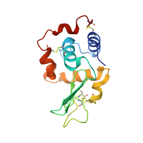
It has been generally believed that polar residues are usually located on the surface of protein structures. However, there are many polar groups in the interior of the structures in reality. To evaluate the contribution of such buried polar groups to the conformational stability of a protein, nonpolar to polar mutations (L8T, A9S, A32S, I56T, I59T, I59S, A92S, V93T, A96S, V99T, and V100T) in the interior of a human lysozyme were examined. The thermodynamic parameters for denaturation were determined using a differential scanning calorimeter, and the crystal structures were analyzed by X-ray crystallography. If a polar group had a heavy energy cost to be buried, a mutant protein would be remarkably destabilized. However, the stability (Delta G) of the Ala to Ser and Val to Thr mutant human lysozymes was comparable to that of the wild-type protein, suggesting a low-energy penalty of buried polar groups. The structural analysis showed that all polar side chains introduced in the mutant proteins were able to find their hydrogen bond partners, which are ubiquitous in protein structures. The empirical structure-based calculation of stability change (Delta Delta G) [Takano et al. (1999) Biochemistry 38, 12698--12708] revealed that the mutant proteins decreased the hydrophobic effect contributing to the stability (Delta G(HP)), but this destabilization was recovered by the hydrogen bonds newly introduced. The present study shows the favorable contribution of polar groups with hydrogen bonds in the interior of protein molecules to the conformational stability.
- Institute for Protein Research, Osaka University, Yamadaoka, Suita, Osaka 565-0871, Japan.
Organizational Affiliation: