1G1G

CRYSTAL STRUCTURE OF PROTEIN TYROSINE PHOSPHATASE 1B COMPLEXED WITH A MONO-PHOSPHORYLATED PEPTIDE (ETDY(PTR)RKGGKGLL) FROM THE INSULIN RECEPTOR KINASE


Experimental Data Snapshot

  • Method: X-RAY DIFFRACTION
  • Resolution: 2.20 Å
  • R-Value Free: 0.229 
  • R-Value Work: 0.212 

wwPDB Validation   3D Report Full Report


This is version 1.3 of the entry. See complete history


Literature

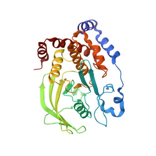
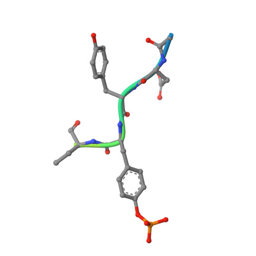
Molecular basis for the dephosphorylation of the activation segment of the insulin receptor by protein tyrosine phosphatase 1B.

Salmeen, A.Andersen, J.N.Myers, M.P.Tonks, N.K.Barford, D.

(2000) Mol Cell 6: 1401-1412

  • DOI: https://doi.org/10.1016/s1097-2765(00)00137-4
  • Primary Citation of Related Structures:  
    1G1F, 1G1G, 1G1H

  • PubMed Abstract: 

    The protein tyrosine phosphatase PTP1B is responsible for negatively regulating insulin signaling by dephosphorylating the phosphotyrosine residues of the insulin receptor kinase (IRK) activation segment. Here, by integrating crystallographic, kinetic, and PTP1B peptide binding studies, we define the molecular specificity of this reaction. Extensive interactions are formed between PTP1B and the IRK sequence encompassing the tandem pTyr residues at 1162 and 1163 such that pTyr-1162 is selected at the catalytic site and pTyr-1163 is located within an adjacent pTyr recognition site. This selectivity is attributed to the 70-fold greater affinity for tandem pTyr-containing peptides relative to mono-pTyr peptides and predicts a hierarchical dephosphorylation process. Many elements of the PTP1B-IRK interaction are unique to PTP1B, indicating that it may be feasible to generate specific, small molecule inhibitors of this interaction to treat diabetes and obesity.


  • Organizational Affiliation

    Laboratory of Molecular Biophysics, University of Oxford, South Parks Road, Oxford, OX1 3QU, United Kingdom.


Macromolecules
Find similar proteins by:  (by identity cutoff)  |  3D Structure
Entity ID: 1
MoleculeChains Sequence LengthOrganismDetailsImage
PROTEIN TYROSINE PHOSPHATASE 1B298Homo sapiensMutation(s): 1 
EC: 3.1.3.48
UniProt & NIH Common Fund Data Resources
Find proteins for P18031 (Homo sapiens)
Explore P18031 
Go to UniProtKB:  P18031
PHAROS:  P18031
GTEx:  ENSG00000196396 
Entity Groups  
Sequence Clusters30% Identity50% Identity70% Identity90% Identity95% Identity100% Identity
UniProt GroupP18031
Sequence Annotations
Expand
  • Reference Sequence

Find similar proteins by:  Sequence   |   3D Structure  

Entity ID: 2
MoleculeChains Sequence LengthOrganismDetailsImage
MONO-PHOSPHORYLATED PEPTIDE FROM THE INSULIN RECEPTOR KINASE13N/AMutation(s): 0 
Entity Groups  
Sequence Clusters30% Identity50% Identity70% Identity90% Identity95% Identity100% Identity
Sequence Annotations
Expand
  • Reference Sequence
Small Molecules
Modified Residues  1 Unique
IDChains TypeFormula2D DiagramParent
PTR
Query on PTR
B
L-PEPTIDE LINKINGC9 H12 N O6 PTYR
Experimental Data & Validation

Experimental Data

  • Method: X-RAY DIFFRACTION
  • Resolution: 2.20 Å
  • R-Value Free: 0.229 
  • R-Value Work: 0.212 
  • Space Group: P 1 21 1
Unit Cell:
Length ( Å )Angle ( ˚ )
a = 44.492α = 90
b = 89.281β = 99.73
c = 52.922γ = 90
Software Package:
Software NamePurpose
AMoREphasing
CNSrefinement
DENZOdata reduction
SCALEPACKdata scaling

Structure Validation

View Full Validation Report


Entry History 

Deposition Data

Revision History  (Full details and data files)

  • Version 1.0: 2001-01-17
    Type: Initial release
  • Version 1.1: 2008-04-27
    Changes: Version format compliance
  • Version 1.2: 2011-07-13
    Changes: Version format compliance
  • Version 1.3: 2021-11-03
    Changes: Database references, Derived calculations