1FTW

STRUCTURES OF GLYCOGEN PHOSPHORYLASE-INHIBITOR COMPLEXES AND THE IMPLICATIONS FOR STRUCTURE-BASED DRUG DESIGN


Experimental Data Snapshot

  • Method: X-RAY DIFFRACTION
  • Resolution: 2.36 Å
  • R-Value Free: 0.214 
  • R-Value Work: 0.174 

wwPDB Validation   3D Report Full Report


This is version 1.4 of the entry. See complete history


Literature

Kinetic and crystallographic studies of glucopyranose spirohydantoin and glucopyranosylamine analogs inhibitors of glycogen phosphorylase.

Watson, K.A.Chrysina, E.D.Tsitsanou, K.E.Zographos, S.E.Archontis, G.Fleet, G.W.Oikonomakos, N.G.

(2005) Proteins 61: 966-983

  • DOI: https://doi.org/10.1002/prot.20653
  • Primary Citation of Related Structures:  
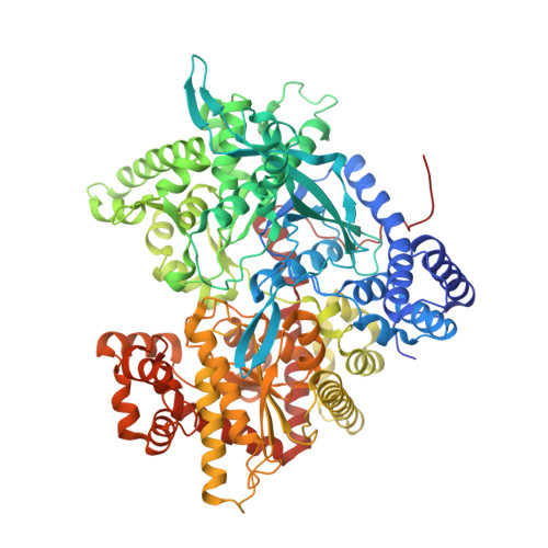
    1FS4, 1FTQ, 1FTW, 1FTY, 1FU4, 1FU7, 1FU8

  • PubMed Abstract: 

    Glycogen phosphorylase (GP) is currently exploited as a target for inhibition of hepatic glycogenolysis under high glucose conditions. Spirohydantoin of glucopyranose and N-acetyl-beta-D-glucopyranosylamine have been identified as the most potent inhibitors of GP that bind at the catalytic site. Four spirohydantoin and three beta-D-glucopyranosylamine analogs have been designed, synthesized and tested for inhibition of GP in kinetic experiments. Depending on the functional group introduced, the K(i) values varied from 16.5 microM to 1200 microM. In order to rationalize the kinetic results, we determined the crystal structures of the analogs in complex with GP. All the inhibitors bound at the catalytic site of the enzyme, by making direct and water-mediated hydrogen bonds with the protein and by inducing minor movements of the side chains of Asp283 and Asn284, of the 280s loop that blocks access of the substrate glycogen to the catalytic site, and changes in the water structure in the vicinity of the site. The differences observed in the Ki values of the analogs can be interpreted in terms of variations in hydrogen bonding and van der Waals interactions, desolvation effects, ligand conformational entropy, and displacement of water molecules on ligand binding to the catalytic site.


  • Organizational Affiliation

    Laboratory of Molecular Biophysics, University of Oxford, Oxford, United Kingdom.


Macromolecules
Find similar proteins by:  (by identity cutoff)  |  3D Structure
Entity ID: 1
MoleculeChains Sequence LengthOrganismDetailsImage
GLYCOGEN PHOSPHORYLASE842Oryctolagus cuniculusMutation(s): 0 
EC: 2.4.1.1
UniProt
Find proteins for P00489 (Oryctolagus cuniculus)
Explore P00489 
Go to UniProtKB:  P00489
Entity Groups  
Sequence Clusters30% Identity50% Identity70% Identity90% Identity95% Identity100% Identity
UniProt GroupP00489
Sequence Annotations
Expand
  • Reference Sequence
Small Molecules
Ligands 2 Unique
IDChains Name / Formula / InChI Key2D Diagram3D Interactions
GL5
Query on GL5

Download Ideal Coordinates CCD File 
B [auth A](5S,7R,8S,9S,10R)-3,8,9,10-tetrahydroxy-7-(hydroxymethyl)-6-oxa-1,3-diazaspiro[4.5]decane-2,4-dione
C8 H12 N2 O8
IHDZZZHACPJCFJ-WWHASAIZSA-N
PLP
Query on PLP

Download Ideal Coordinates CCD File 
C [auth A]PYRIDOXAL-5'-PHOSPHATE
C8 H10 N O6 P
NGVDGCNFYWLIFO-UHFFFAOYSA-N
Binding Affinity Annotations 
IDSourceBinding Affinity
GL5 BindingDB:  1FTW Ki: 3.89e+4 (nM) from 1 assay(s)
Experimental Data & Validation

Experimental Data

  • Method: X-RAY DIFFRACTION
  • Resolution: 2.36 Å
  • R-Value Free: 0.214 
  • R-Value Work: 0.174 
  • Space Group: P 43 21 2
Unit Cell:
Length ( Å )Angle ( ˚ )
a = 128.5α = 90
b = 128.5β = 90
c = 116.3γ = 90
Software Package:
Software NamePurpose
CCP4model building
X-PLORrefinement
XDSdata reduction
XDSdata scaling
CCP4phasing

Structure Validation

View Full Validation Report


Entry History 

Deposition Data

Revision History  (Full details and data files)

  • Version 1.0: 2000-10-04
    Type: Initial release
  • Version 1.1: 2008-04-27
    Changes: Version format compliance
  • Version 1.2: 2011-07-13
    Changes: Derived calculations, Version format compliance
  • Version 1.3: 2020-07-29
    Type: Remediation
    Reason: Carbohydrate remediation
    Changes: Derived calculations, Structure summary
  • Version 1.4: 2023-08-09
    Changes: Data collection, Database references, Refinement description, Structure summary