1FIP

THE STRUCTURE OF FIS MUTANT PRO61ALA ILLUSTRATES THAT THE KINK WITHIN THE LONG ALPHA-HELIX IS NOT DUE TO THE PRESENCE OF THE PROLINE RESIDUE


Experimental Data Snapshot

  • Method: X-RAY DIFFRACTION
  • Resolution: 1.90 Å
  • R-Value Work: 0.199 
  • R-Value Observed: 0.199 

wwPDB Validation   3D Report Full Report


This is version 1.4 of the entry. See complete history


Literature

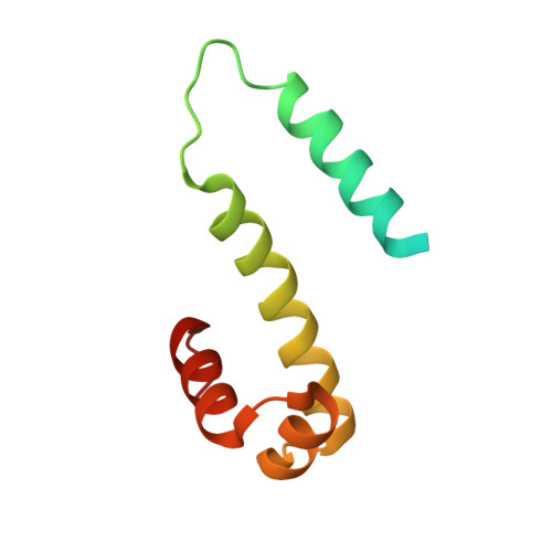
The structure of Fis mutant Pro61Ala illustrates that the kink within the long alpha-helix is not due to the presence of the proline residue.

Yuan, H.S.Wang, S.S.Yang, W.Z.Finkel, S.E.Johnson, R.C.

(1994) J Biol Chem 269: 28947-28954

  • DOI: https://doi.org/10.2210/pdb1fip/pdb
  • Primary Citation of Related Structures:  
    1FIP

  • PubMed Abstract: 

    The influence of proline on bending of the alpha-helix was investigated by replacement of the proline residue located in the middle of the long alpha-helix of the Fis protein with alanine, serine, or leucine. Each of the three substitutions folded into a stable protein with the same or higher melting points than the wild-type, but only Pro61Ala was functionally active in stimulating Hin-mediated DNA inversion. Pro61Ala formed crystals that were isomorphous with the wild-type protein allowing the structure to be determined at 1.9-A resolution by x-ray diffraction methods. The structure of the Pro61Ala mutant is almost identical to the wild-type protein, consistent with its near wild-type activity. One of the alpha-helices, the B-helix, is kinked in the wild-type Fis protein by 20 degrees which was previously assumed to be caused solely by the presence of proline 61 in the center of the helix. However, the B-helix is still kinked by 16 degrees when proline 61 is replaced by alanine. Local peptide backbone movement around residue 57 adjusts the geometry of the helix to accommodate the new main chain hydrogen bond between the -CO group in Glu57 and the -NH group in Ala61. Thus, the kink of the alpha-helix in Pro61Ala does not require the presence of proline.


  • Organizational Affiliation

    Institute of Molecular Biology, Academia Sinica, Taipei, Taiwan, Republic of China.


Macromolecules
Find similar proteins by:  (by identity cutoff)  |  3D Structure
Entity ID: 1
MoleculeChains Sequence LengthOrganismDetailsImage
FACTOR FOR INVERSION STIMULATION (FIS)
A, B
98Escherichia coliMutation(s): 1 
Gene Names: FIS
UniProt
Find proteins for P0A6R3 (Escherichia coli (strain K12))
Explore P0A6R3 
Go to UniProtKB:  P0A6R3
Entity Groups  
Sequence Clusters30% Identity50% Identity70% Identity90% Identity95% Identity100% Identity
UniProt GroupP0A6R3
Sequence Annotations
Expand
  • Reference Sequence

Find similar proteins by:  Sequence   |   3D Structure  

Entity ID: 2
MoleculeChains Sequence LengthOrganismDetailsImage
UNKNOWN PEPTIDE, POSSIBLY PART OF THE UNOBSERVED RESIDUES IN ENTITY 1
C, D
4N/AMutation(s): 0 
Sequence Annotations
Expand
  • Reference Sequence
Experimental Data & Validation

Experimental Data

  • Method: X-RAY DIFFRACTION
  • Resolution: 1.90 Å
  • R-Value Work: 0.199 
  • R-Value Observed: 0.199 
  • Space Group: P 21 21 21
Unit Cell:
Length ( Å )Angle ( ˚ )
a = 79.08α = 90
b = 51.12β = 90
c = 47.29γ = 90
Software Package:
Software NamePurpose
X-PLORmodel building
X-PLORrefinement
X-PLORphasing

Structure Validation

View Full Validation Report


Entry History 

Deposition Data

Revision History  (Full details and data files)

  • Version 1.0: 1995-02-14
    Type: Initial release
  • Version 1.1: 2008-03-24
    Changes: Version format compliance
  • Version 1.2: 2011-07-13
    Changes: Version format compliance
  • Version 1.3: 2015-08-05
    Changes: Structure summary
  • Version 1.4: 2024-02-07
    Changes: Data collection, Database references, Other, Structure summary