1FAZ | pdb_00001faz

THE CRYSTAL STRUCTURE OF PROKARYOTIC PHOSPHOLIPASE A2


Experimental Data Snapshot

  • Method: X-RAY DIFFRACTION
  • Resolution: 1.40 Å
  • R-Value Free: 
    0.231 (Depositor) 
  • R-Value Work: 
    0.188 (Depositor) 
  • R-Value Observed: 
    0.188 (Depositor) 

wwPDB Validation 3D Report Full Report

Validation slider image for 1FAZ

This is version 1.4 of the entry. See complete history

Literature

The crystal structure of prokaryotic phospholipase A2.

Matoba, Y.Katsube, Y.Sugiyama, M.

(2002) J Biological Chem 277: 20059-20069

  • DOI: https://doi.org/10.1074/jbc.M200263200
  • Primary Citation Related Structures: 
    1FAZ, 1KP4

  • PubMed Abstract: 

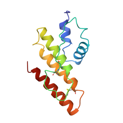
    In this study, the x-ray crystal structures of the calcium-free and calcium-bound forms of phospholipase A(2) (PLA(2)), produced extracellularly by Streptomyces violaceoruber, were determined by using the multiple isomorphous replacement and molecular replacement methods, respectively. The former and latter structures were refined to an R-factor of 18.8% at a 1.4-A resolution and an R-factor of 15.0% at a 1.6-A resolution, respectively. The overall structure of the prokaryotic PLA(2) exhibits a novel folding topology that demonstrates that it is completely distinct from those of eukaryotic PLA(2)s, which have been already determined by x-ray and NMR analyses. Furthermore, the coordination geometry of the calcium(II) ion apparently deviated from that of eukaryotic PLA(2)s. Regardless of the evolutionary divergence, the catalytic mechanism including the calcium(II) ion on secreted PLA(2) seems to be conserved between prokaryotic and eukaryotic cells. Demonstrating that the overall structure determined by x-ray analysis is almost the same as that determined by NMR analysis is useful to discuss the catalytic mechanism at the molecular level of the bacterial PLA(2).


  • Organizational Affiliation
    • Institute of Pharmaceutical Sciences, Faculty of Medicine, Hiroshima University, Kasumi 1-2-3, Minami-ku, Hiroshima 734-8551, Japan.

Macromolecule Content 

  • Total Structure Weight: 13.57 kDa 
  • Atom Count: 1,085 
  • Modeled Residue Count: 122 
  • Deposited Residue Count: 122 
  • Unique protein chains: 1

Macromolecules

Find similar proteins by:|  3D Structure
Entity ID: 1
MoleculeChains  Sequence LengthOrganismDetailsImage
PHOSPHOLIPASE A2122Streptomyces violaceoruberMutation(s): 0 
EC: 3.1.1.4
UniProt
Find proteins for Q6UV28 (Streptomyces violaceoruber)
Explore Q6UV28 
Go to UniProtKB:  Q6UV28
Entity Groups
Sequence Clusters30% Identity50% Identity70% Identity90% Identity95% Identity100% Identity
UniProt GroupQ6UV28
Sequence Annotations
Expand
Reference Sequence

Experimental Data & Validation

Experimental Data

  • Method: X-RAY DIFFRACTION
  • Resolution: 1.40 Å
  • R-Value Free:  0.231 (Depositor) 
  • R-Value Work:  0.188 (Depositor) 
  • R-Value Observed: 0.188 (Depositor) 
Space Group: P 1 21 1
Unit Cell:
Length ( Å )Angle ( ˚ )
a = 29.41α = 90
b = 57.75β = 111.48
c = 31.71γ = 90
Software Package:
Software NamePurpose
PHASESphasing
X-PLORrefinement

Structure Validation

View Full Validation Report


Entry History 

Deposition Data

Revision History  (Full details and data files)

  • Version 1.0: 2001-07-18
    Type: Initial release
  • Version 1.1: 2008-04-27
    Changes: Version format compliance
  • Version 1.2: 2011-07-13
    Changes: Version format compliance
  • Version 1.3: 2017-10-04
    Changes: Refinement description
  • Version 1.4: 2024-11-20
    Changes: Data collection, Database references, Structure summary