1F5N

HUMAN GUANYLATE BINDING PROTEIN-1 IN COMPLEX WITH THE GTP ANALOGUE, GMPPNP.


Experimental Data Snapshot

  • Method: X-RAY DIFFRACTION
  • Resolution: 1.70 Å
  • R-Value Free: 0.255 
  • R-Value Work: 0.226 
  • R-Value Observed: 0.226 

wwPDB Validation   3D Report Full Report


This is version 1.5 of the entry. See complete history


Literature

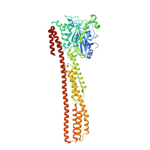
Triphosphate structure of guanylate-binding protein 1 and implications for nucleotide binding and GTPase mechanism.

Prakash, B.Renault, L.Praefcke, G.J.Herrmann, C.Wittinghofer, A.

(2000) EMBO J 19: 4555-4564

  • DOI: https://doi.org/10.1093/emboj/19.17.4555
  • Primary Citation of Related Structures:  
    1F5N

  • PubMed Abstract: 
  • The interferon-gamma-induced guanylate-binding protein 1 (GBP1) belongs to a special class of large GTP- binding proteins of 60-100 kDa with unique characteristics. Here we present the structure of human GBP1 in complex with the non-hydrolysable GTP analogue GppNHp ...

    The interferon-gamma-induced guanylate-binding protein 1 (GBP1) belongs to a special class of large GTP- binding proteins of 60-100 kDa with unique characteristics. Here we present the structure of human GBP1 in complex with the non-hydrolysable GTP analogue GppNHp. Basic features of guanine nucleotide binding, such as the P-loop orientation and the Mg(2+) co-ordination, are analogous to those of Ras-related and heterotrimeric GTP-binding proteins. However, the glycosidic bond and thus the orientation of the guanine base and its interaction with the protein are very different. Furthermore, two unique regions around the base and the phosphate-binding areas, the guanine and the phosphate caps, respectively, give the nucleotide-binding site a unique appearance not found in the canonical GTP-binding proteins. The phosphate cap, which constitutes the region analogous to switch I, completely shields the phosphate-binding site from solvent such that a potential GTPase-activating protein cannot approach. This has consequences for the GTPase mechanism of hGBP1 and possibly of other large GTP-binding proteins.


    Related Citations: 
    • Structure of human Guanylate Binding Protein-1 representing a unique class of GTP binding proteins
      Prakash, B., Praefcke, G.J.K., Renault, L., Wittinghofer, A., Herrmann, C.
      (2000) Nature 403: 567
    • Nucleotide-binding Characteristics of Human Guanylate-binding protein 1(hGBP1) and Identification of the Third GTP-binding Motif
      Praefcke, G.J.K., Geyer, M., Schwemmle, M., Kalbitzer, H.R., Herrmann, C.
      (1999) J Mol Biol 292: 321

    Organizational Affiliation

    Max-Planck-Institut für molekulare Physiologie, Otto-Hahn-Strabetae 11, D-44227 Dortmund, Germany.


Macromolecules
Find similar proteins by:  (by identity cutoff)  |  3D Structure
Entity ID: 1
MoleculeChainsSequence LengthOrganismDetailsImage
INTERFERON-INDUCED GUANYLATE-BINDING PROTEIN 1592Homo sapiensMutation(s): 0 
Gene Names: GBP1
EC: 3.6.5
UniProt & NIH Common Fund Data Resources
Find proteins for P32455 (Homo sapiens)
Explore P32455 
Go to UniProtKB:  P32455
PHAROS:  P32455
Entity Groups  
Sequence Clusters30% Identity50% Identity70% Identity90% Identity95% Identity100% Identity
UniProt GroupP32455
Protein Feature View
Expand
  • Reference Sequence
Small Molecules
Ligands 2 Unique
IDChainsName / Formula / InChI Key2D Diagram3D Interactions
GNP
Query on GNP

Download Ideal Coordinates CCD File 
C [auth A]PHOSPHOAMINOPHOSPHONIC ACID-GUANYLATE ESTER
C10 H17 N6 O13 P3
UQABYHGXWYXDTK-UUOKFMHZSA-N
 Ligand Interaction
MG
Query on MG

Download Ideal Coordinates CCD File 
B [auth A]MAGNESIUM ION
Mg
JLVVSXFLKOJNIY-UHFFFAOYSA-N
 Ligand Interaction
Experimental Data & Validation

Experimental Data

  • Method: X-RAY DIFFRACTION
  • Resolution: 1.70 Å
  • R-Value Free: 0.255 
  • R-Value Work: 0.226 
  • R-Value Observed: 0.226 
  • Space Group: C 1 2 1
Unit Cell:
Length ( Å )Angle ( ˚ )
a = 161.228α = 90
b = 42.652β = 100.19
c = 91.544γ = 90
Software Package:
Software NamePurpose
AMoREphasing
CNSrefinement
MAR345data collection
XDSdata scaling

Structure Validation

View Full Validation Report


Entry History 

Deposition Data

Revision History  (Full details and data files)

  • Version 1.0: 2000-09-27
    Type: Initial release
  • Version 1.1: 2008-04-27
    Changes: Version format compliance
  • Version 1.2: 2011-07-13
    Changes: Version format compliance
  • Version 1.3: 2017-10-04
    Changes: Refinement description
  • Version 1.4: 2018-01-31
    Changes: Experimental preparation
  • Version 1.5: 2023-08-09
    Changes: Data collection, Database references, Derived calculations, Refinement description