1EVH

EVH1 DOMAIN FROM MURINE ENABLED IN COMPLEX WITH ACTA PEPTIDE


Experimental Data Snapshot

  • Method: X-RAY DIFFRACTION
  • Resolution: 1.80 Å
  • R-Value Free: 0.268 
  • R-Value Work: 0.230 

wwPDB Validation   3D Report Full Report


This is version 1.3 of the entry. See complete history


Literature

Structure of the enabled/VASP homology 1 domain-peptide complex: a key component in the spatial control of actin assembly.

Prehoda, K.E.Lee, D.J.Lim, W.A.

(1999) Cell 97: 471-480

  • DOI: https://doi.org/10.1016/s0092-8674(00)80757-6
  • Primary Citation of Related Structures:  
    1EVH

  • PubMed Abstract: 

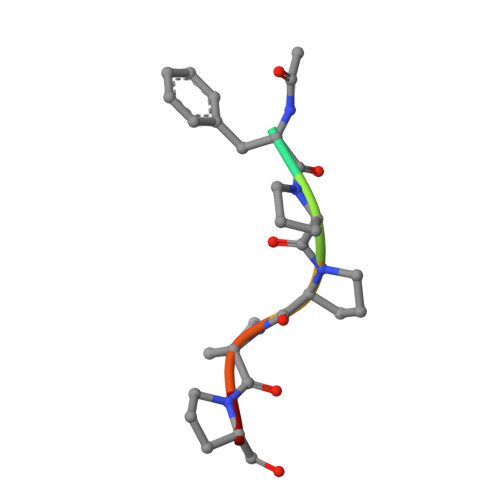
    The Enabled/VASP homology 1 (EVH1; also called WH1) domain is an interaction module found in several proteins implicated in actin-based cell motility. EVH1 domains bind the consensus proline-rich motif FPPPP and are required for targeting the actin assembly machinery to sites of cytoskeletal remodeling. The crystal structure of the mammalian Enabled (Mena) EVH1 domain complexed with a peptide ligand reveals a mechanism of recognition distinct from that used by other proline-binding modules. The EVH1 domain fold is unexpectedly similar to that of the pleckstrin homology domain, a membrane localization module. This finding demonstrates the functional plasticity of the pleckstrin homology fold as a binding scaffold and suggests that membrane association may play an auxiliary role in EVH1 targeting.


  • Organizational Affiliation

    Department of Cellular and Molecular Pharmacology, University of California, San Francisco 94143, USA.


Macromolecules
Find similar proteins by:  (by identity cutoff)  |  3D Structure
Entity ID: 1
MoleculeChains Sequence LengthOrganismDetailsImage
PROTEIN (MENA EVH1 DOMAIN)112Mus musculusMutation(s): 0 
UniProt
Find proteins for Q03173 (Mus musculus)
Explore Q03173 
Go to UniProtKB:  Q03173
Entity Groups  
Sequence Clusters30% Identity50% Identity70% Identity90% Identity95% Identity100% Identity
UniProt GroupQ03173
Sequence Annotations
Expand
  • Reference Sequence

Find similar proteins by:  Sequence   |   3D Structure  

Entity ID: 2
MoleculeChains Sequence LengthOrganismDetailsImage
Peptide ACTA7N/AMutation(s): 0 
Sequence Annotations
Expand
  • Reference Sequence
Experimental Data & Validation

Experimental Data

  • Method: X-RAY DIFFRACTION
  • Resolution: 1.80 Å
  • R-Value Free: 0.268 
  • R-Value Work: 0.230 
  • Space Group: C 2 2 21
Unit Cell:
Length ( Å )Angle ( ˚ )
a = 33.614α = 90
b = 62.566β = 90
c = 94.316γ = 90
Software Package:
Software NamePurpose
SHARPphasing
CNSrefinement
DENZOdata reduction
SCALEPACKdata scaling

Structure Validation

View Full Validation Report


Entry History 

Deposition Data

Revision History  (Full details and data files)

  • Version 1.0: 1999-05-21
    Type: Initial release
  • Version 1.1: 2008-04-26
    Changes: Version format compliance
  • Version 1.2: 2011-07-13
    Changes: Version format compliance
  • Version 1.3: 2023-12-27
    Changes: Data collection, Database references, Derived calculations