Mutation of the iron ligand His 249 to Glu in the N-lobe of human transferrin abolishes the dilysine "trigger" but does not significantly affect iron release.
MacGillivray, R.T., Bewley, M.C., Smith, C.A., He, Q.Y., Mason, A.B.(2000) Biochemistry 39: 1211-1216
- PubMed: 10684598 Search on PubMed
- DOI: https://doi.org/10.1021/bi991522y
- Primary Citation Related Structures:
1DTG - PubMed Abstract:
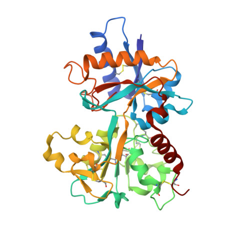
Serum transferrin is the major iron transport protein in humans. Its function depends on its ability to bind iron with very high affinity, yet to release this bound iron at the lower intracellular pH. Possible explanations for the release of iron from transferrin at low pH include protonation of a histidine ligand and the existence of a pH-sensitive "trigger" involving a hydrogen-bonded pair of lysines in the N-lobe of transferrin. We have determined the crystal structure of the His249Glu mutant of the N-lobe half-molecule of human transferrin and compared its iron-binding properties with those of the wild-type protein and other mutants. The crystal structure, determined at 2.4 A resolution (R-factor 19.8%, R(free) 29.4%), shows that Glu 249 is directly bound to iron, in place of the His ligand, and that a local movement of Lys 296 has broken the dilysine interaction. Despite the loss of this potentially pH-sensitive interaction, the H249E mutant is only slightly more acid-stable than wild-type and releases iron slightly faster. We conclude that the loss of the dilysine interaction does make the protein more acid stable but that this is counterbalanced by the replacement of a neutral ligand (His) by a negatively charged one (Glu), thus disrupting the electroneutrality of the binding site.
- Department of Biochemistry, Massey University, Palmerston North, New Zealand.
Organizational Affiliation: