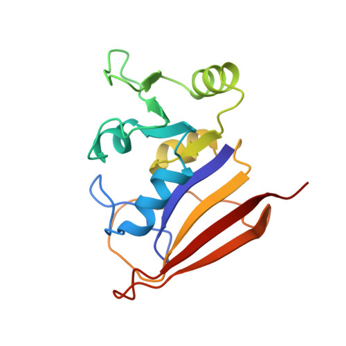
Solution structure of a brodimoprim analogue in its complex with Lactobacillus casei dihydrofolate reductase.
Morgan, W.D., Birdsall, B., Polshakov, V.I., Sali, D., Kompis, I., Feeney, J.(1995) Biochemistry 34: 11690-11702
- PubMed: 7547901 Search on PubMed
- DOI: https://doi.org/10.1021/bi00037a006
- Primary Citation Related Structures:
1DIS, 1DIU - PubMed Abstract:
Two-dimensional (2D) double-quantum-filtered correlation spectroscopy (DQF-COSY), total correlation spectroscopy (TOCSY), nuclear Overhauser effect spectroscopy (NOESY), and rotating-frame NOESY (ROESY) spectra were used to assign essentially all the protons in a 1:1 complex of Lactobacillus casei dihydrofolate reductase formed with an analogue of the antibacterial drug brodimoprim [2,4-diamino-5-(3',5'-dimethoxy-4'-bromobenzyl)pyrimidine]. The analogue has a 4,6-dicarboxylic acid side chain substituted on the 3'-O position designed to interact with the Arg 57 and His 28 residues in L. casei dihydrofolate reductase; it binds a factor of 10(3) more tightly to the enzyme than does the parent compound. Thirty-eight intermolecular and 11 intramolecular NOEs were measured involving the bound brodimoprim-4,6-dicarboxylic acid analogue. These provided the distance constraints used in conjunction with an energy minimization and simulated annealing protocol (using Discover from Biosym Ltd.) to dock the brodimoprim analogue into dihydrofolate reductase. In calculations where side chains and backbone fragments for binding-site residues were allowed flexibility, 90% of the 40 calculated structures had reasonable covalent geometry and none of them had NOE distance violations of greater than 0.36 A. The conformations of the aromatic rings in the bound ligand were well-defined in all the structures, with torsion angles tau 1 = -153 degrees +/- 4 degrees (C4-C5-C7-C1') and tau 2 = 53 degrees +/- 4 degrees (C5-C7-C1'-C2'): the aromatic rings of the ligand occupied essentially the same space in all the calculated structures (root mean square deviation value 1.83 A). Inclusion of the electrostatic interactions into the energy minimizations indicated that structures in which the 4,6-dicarboxylate group of the ligand interacts with the side chains of Arg 57 and His 28 are of low energy. Significant differences in side-chain and backbone conformations were detected between binding-site residues in the enzyme complexes with the brodimorpim analogue and methotrexate.
- Laboratory of Molecular Structure, National Institute for Medical Research, Mill Hill, London, U.K.
Organizational Affiliation: