1D2E

CRYSTAL STRUCTURE OF MITOCHONDRIAL EF-TU IN COMPLEX WITH GDP


Experimental Data Snapshot

  • Method: X-RAY DIFFRACTION
  • Resolution: 1.94 Å
  • R-Value Free: 0.257 
  • R-Value Work: 0.237 
  • R-Value Observed: 0.237 

wwPDB Validation   3D Report Full Report


Ligand Structure Quality Assessment 


This is version 1.4 of the entry. See complete history


Literature

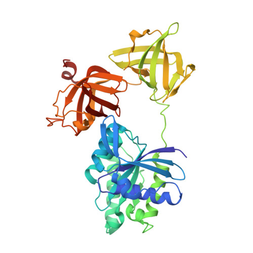
High resolution crystal structure of bovine mitochondrial EF-Tu in complex with GDP.

Andersen, G.R.Thirup, S.Spremulli, L.L.Nyborg, J.

(2000) J Mol Biol 297: 421-436

  • DOI: https://doi.org/10.1006/jmbi.2000.3564
  • Primary Citation of Related Structures:  
    1D2E

  • PubMed Abstract: 

    The crystal structure of bovine mitochondrial elongation factor Tu (EF-Tu) in complex with GDP has been determined at a resolution of 1. 94 A. The structure is similar to that of EF-Tu:GDP from Escherichia coli and Thermus aquaticus, but the orientation of the GDP-binding domain 1 is changed relative to domains 2 and 3. Sixteen conserved water molecules common to EF-Tu and other G-proteins in the GDP-binding site are described. These water molecules create a network linking separated parts of the binding pocket. Mitochondrial EF-Tu binds nucleotides less tightly than prokaryotic EF-Tu possibly due to an increased mobility in regions close to the GDP-binding site. The C-terminal extension of mitochondrial EF-Tu has structural similarities with DNA recognising zinc fingers suggesting that the extension may be involved in recognition of RNA.


  • Organizational Affiliation

    Institute of Molecular and Structural Biology, Aarhus University, Gustav Wiedsvej 10C, Aarhus, DK8000, Denmark.


Macromolecules
Find similar proteins by:  (by identity cutoff)  |  3D Structure
Entity ID: 1
MoleculeChains Sequence LengthOrganismDetailsImage
ELONGATION FACTOR TU (EF-TU)
A, B, C, D
397Bos taurusMutation(s): 0 
UniProt
Find proteins for P49410 (Bos taurus)
Explore P49410 
Go to UniProtKB:  P49410
Entity Groups  
Sequence Clusters30% Identity50% Identity70% Identity90% Identity95% Identity100% Identity
UniProt GroupP49410
Sequence Annotations
Expand
  • Reference Sequence
Small Molecules
Experimental Data & Validation

Experimental Data

  • Method: X-RAY DIFFRACTION
  • Resolution: 1.94 Å
  • R-Value Free: 0.257 
  • R-Value Work: 0.237 
  • R-Value Observed: 0.237 
  • Space Group: P 1 21 1
Unit Cell:
Length ( Å )Angle ( ˚ )
a = 59.09α = 90
b = 119.78β = 96.97
c = 128.89γ = 90
Software Package:
Software NamePurpose
AMoREphasing
CNSrefinement
DENZOdata reduction
SCALEPACKdata scaling

Structure Validation

View Full Validation Report


Ligand Structure Quality Assessment 


Entry History 

Deposition Data

Revision History  (Full details and data files)

  • Version 1.0: 1999-09-28
    Type: Initial release
  • Version 1.1: 2008-04-27
    Changes: Version format compliance
  • Version 1.2: 2011-07-13
    Changes: Version format compliance
  • Version 1.3: 2018-01-31
    Changes: Experimental preparation
  • Version 1.4: 2024-02-07
    Changes: Data collection, Database references, Derived calculations