1D0Z

DICTYOSTELIUM MYOSIN S1DC (MOTOR DOMAIN FRAGMENT) COMPLEXED WITH P-NITROPHENYL AMINOETHYLDIPHOSPHATE BERYLLIUM TRIFLUORIDE.


Experimental Data Snapshot

  • Method: X-RAY DIFFRACTION
  • Resolution: 2.00 Å
  • R-Value Work: 0.193 
  • R-Value Observed: 0.193 

wwPDB Validation   3D Report Full Report


Ligand Structure Quality Assessment 


This is version 1.5 of the entry. See complete history


Literature

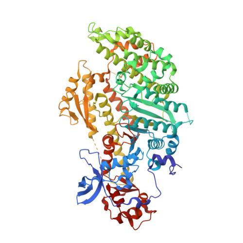
X-ray structures of the Dictyostelium discoideum myosin motor domain with six non-nucleotide analogs.

Gulick, A.M.Bauer, C.B.Thoden, J.B.Pate, E.Yount, R.G.Rayment, I.

(2000) J Biol Chem 275: 398-408

  • DOI: https://doi.org/10.1074/jbc.275.1.398
  • Primary Citation of Related Structures:  
    1D0X, 1D0Y, 1D0Z, 1D1A, 1D1B, 1D1C

  • PubMed Abstract: 

    The three-dimensional structures of the truncated myosin head from Dictyostelium discoideum myosin II complexed with dinitrophenylaminoethyl-, dinitrophenylaminopropyl-, o-nitrophenylaminoethyl-, m-nitrophenylaminoethyl-, p-nitrophenylaminoethyl-, and o-nitrophenyl-N-methyl-aminoethyl-diphosphate.beryllium fluoride have been determined to better than 2.3-A resolution. The structure of the protein and nucleotide binding pocket in these complexes is very similar to that of S1dC.ADP.BeF(x) (Fisher, A. J., Smith, C. A., Thoden, J., Smith, R., Sutoh, K., Holden, H. M., and Rayment, I. (1995) Biochemistry 34, 8960-8972). The position of the triphosphate-like moiety is essentially identical in all complexes. Furthermore, the alkyl-amino group plays the same role as the ribose by linking the triphosphate to the adenine binding pocket; however, none of the phenyl groups lie in the same position as adenine in S1dC.MgADP.BeF(x), even though several of these nucleotide analogs are functionally equivalent to ATP. Rather the former location of adenine is occupied by water in the nanolog complexes, and the phenyl groups are organized in a manner that attempts to optimize their hydrogen bonding interactions with this constellation of solvent molecules. A comparison of the kinetic and structural properties of the nanologs relative to ATP suggests that the ability of a substrate to sustain tension and to generate movement correlates with a well defined interaction with the active site water structure observed in S1dC.MgADP.BeF(x).


  • Organizational Affiliation

    Institute for Enzyme Research, Department of Biochemistry, University of Wisconsin, Madison, Wisconsin 53705, USA.


Macromolecules
Find similar proteins by:  (by identity cutoff)  |  3D Structure
Entity ID: 1
MoleculeChains Sequence LengthOrganismDetailsImage
MYOSIN761Dictyostelium discoideumMutation(s): 2 
UniProt
Find proteins for P08799 (Dictyostelium discoideum)
Explore P08799 
Go to UniProtKB:  P08799
Entity Groups  
Sequence Clusters30% Identity50% Identity70% Identity90% Identity95% Identity100% Identity
UniProt GroupP08799
Sequence Annotations
Expand
  • Reference Sequence
Small Molecules
Ligands 2 Unique
IDChains Name / Formula / InChI Key2D Diagram3D Interactions
PNQ
Query on PNQ

Download Ideal Coordinates CCD File 
C [auth A]P-NITROPHENYL AMINOETHYLDIPHOSPHATE BERYLLIUM TRIFLUORIDE
C8 H11 Be F3 N2 O9 P2
VQFXKFKMXDVHLH-UHFFFAOYSA-J
MG
Query on MG

Download Ideal Coordinates CCD File 
B [auth A]MAGNESIUM ION
Mg
JLVVSXFLKOJNIY-UHFFFAOYSA-N
Experimental Data & Validation

Experimental Data

  • Method: X-RAY DIFFRACTION
  • Resolution: 2.00 Å
  • R-Value Work: 0.193 
  • R-Value Observed: 0.193 
  • Space Group: P 21 21 2
Unit Cell:
Length ( Å )Angle ( ˚ )
a = 103.987α = 90
b = 180.855β = 90
c = 54.101γ = 90
Software Package:
Software NamePurpose
AMoREphasing
TNTrefinement
DENZOdata reduction
SCALEPACKdata scaling

Structure Validation

View Full Validation Report


Ligand Structure Quality Assessment 


Entry History 

Deposition Data

Revision History  (Full details and data files)

  • Version 1.0: 2000-01-12
    Type: Initial release
  • Version 1.1: 2008-04-27
    Changes: Version format compliance
  • Version 1.2: 2011-07-13
    Changes: Version format compliance
  • Version 1.3: 2018-01-31
    Changes: Experimental preparation
  • Version 1.4: 2021-11-03
    Changes: Database references, Derived calculations
  • Version 1.5: 2024-02-07
    Changes: Data collection