1CXC

CRYSTALLIZATION AND X-RAY STRUCTURE DETERMINATION OF CYTOCHROME C2 FROM RHODOBACTER SPHAEROIDES IN THREE CRYSTAL FORMS


Experimental Data Snapshot

  • Method: X-RAY DIFFRACTION
  • Resolution: 1.60 Å
  • R-Value Observed: 0.225 

wwPDB Validation   3D Report Full Report


Ligand Structure Quality Assessment 


This is version 2.0 of the entry. See complete history


Literature

Crystallization and X-ray structure determination of cytochrome c2 from Rhodobacter sphaeroides in three crystal forms.

Axelrod, H.L.Feher, G.Allen, J.P.Chirino, A.J.Day, M.W.Hsu, B.T.Rees, D.C.

(1994) Acta Crystallogr D Biol Crystallogr 50: 596-602

  • DOI: https://doi.org/10.1107/S0907444994001319
  • Primary Citation of Related Structures:  
    1CXA, 1CXC, 2CXB

  • PubMed Abstract: 

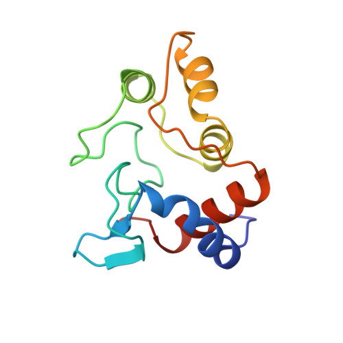
    Cytochrome c(2) serves as the secondary electron donor that reduces the photo-oxidized bacteriochlorophyll dimer in photosynthetic bacteria. Cytochrome c(2) from Rhodobacter sphaeroides has been crystallized in three different forms. At high ionic strength, crystals of a hexagonal space group (P6(1)22) were obtained, while at low ionic strength, triclinic (P1) and tetragonal (P4(1)2(1)2) crystals were formed. The three-dimensional structures of the cytochrome in all three crystal forms have been determined by X-ray diffraction at resolutions of 2.20 A (hexagonal), 1.95 A, (triclinic) and 1.53 A (tetragonal). The most significant difference observed was the binding of an imidazole molecule to the iron atom of the heme group in the hexagonal structure. This binding displaces the sulfur atom of Met l00, which forms the axial ligand in the triclinic and tetragonal structures.


  • Organizational Affiliation

    Department of Physics, University of California, San Diego, La Jolla 92093-0319, USA.


Macromolecules
Find similar proteins by:  (by identity cutoff)  |  3D Structure
Entity ID: 1
MoleculeChains Sequence LengthOrganismDetailsImage
CYTOCHROME C2124Cereibacter sphaeroidesMutation(s): 0 
UniProt
Find proteins for P0C0X8 (Cereibacter sphaeroides)
Explore P0C0X8 
Go to UniProtKB:  P0C0X8
Entity Groups  
Sequence Clusters30% Identity50% Identity70% Identity90% Identity95% Identity100% Identity
UniProt GroupP0C0X8
Sequence Annotations
Expand
  • Reference Sequence
Small Molecules
Ligands 1 Unique
IDChains Name / Formula / InChI Key2D Diagram3D Interactions
HEC
Query on HEC

Download Ideal Coordinates CCD File 
B [auth A]HEME C
C34 H34 Fe N4 O4
HXQIYSLZKNYNMH-LJNAALQVSA-N
Experimental Data & Validation

Experimental Data

  • Method: X-RAY DIFFRACTION
  • Resolution: 1.60 Å
  • R-Value Observed: 0.225 
  • Space Group: P 41 21 2
Unit Cell:
Length ( Å )Angle ( ˚ )
a = 82.33α = 90
b = 82.33β = 90
c = 37.61γ = 90
Software Package:
Software NamePurpose
X-PLORmodel building
TNTrefinement
X-PLORrefinement
X-PLORphasing

Structure Validation

View Full Validation Report


Ligand Structure Quality Assessment 


Entry History 

Deposition Data

Revision History  (Full details and data files)

  • Version 1.0: 1995-09-15
    Type: Initial release
  • Version 1.1: 2008-03-24
    Changes: Version format compliance
  • Version 1.2: 2011-07-13
    Changes: Version format compliance
  • Version 1.3: 2017-11-29
    Changes: Derived calculations, Other
  • Version 2.0: 2021-03-03
    Changes: Atomic model, Data collection, Derived calculations, Non-polymer description, Structure summary