1CW0

CRYSTAL STRUCTURE ANALYSIS OF VERY SHORT PATCH REPAIR (VSR) ENDONUCLEASE IN COMPLEX WITH A DUPLEX DNA


Experimental Data Snapshot

  • Method: X-RAY DIFFRACTION
  • Resolution: 2.30 Å
  • R-Value Free: 0.230 
  • R-Value Work: 0.204 
  • R-Value Observed: 0.204 

wwPDB Validation   3D Report Full Report


This is version 1.3 of the entry. See complete history


Literature

Recognition of a TG mismatch: the crystal structure of very short patch repair endonuclease in complex with a DNA duplex.

Tsutakawa, S.E.Jingami, H.Morikawa, K.

(1999) Cell 99: 615-623

  • DOI: https://doi.org/10.1016/s0092-8674(00)81550-0
  • Primary Citation of Related Structures:  
    1CW0

  • PubMed Abstract: 

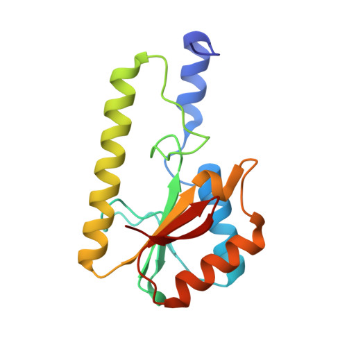
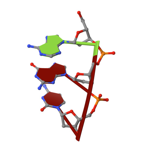
    The crystal structure of very short patch repair (Vsr) endonuclease, in complex with Mg2+ and with duplex DNA containing a TG mismatch, has been determined at 2.3 A resolution. In E. coli, the enzyme recognizes a TG mismatched base pair, generated after spontaneous deamination of methylated cytosines, and cleaves the phosphate backbone on the 5' side of the thymine. Extensive interactions between the DNA and the protein characterize a novel recognition mechanism, where three aromatic residues intercalate from the major groove into the DNA to strikingly deform the base pair stacking. With the presence of a cleaved DNA intermediate in the active center, the structure of the Vsr/DNA complex provides detailed insights into the catalytic mechanism for endonuclease activity.


  • Organizational Affiliation

    Department of Structural Biology, Biomolecular Engineering Research Institute, Suita, Osaka, Japan.


Macromolecules

Find similar proteins by:  (by identity cutoff)  |  3D Structure
Entity ID: 4
MoleculeChains Sequence LengthOrganismDetailsImage
PROTEIN (DNA MISMATCH ENDONUCLEASE)D [auth A]155Escherichia coliMutation(s): 0 
EC: 3.1
UniProt
Find proteins for P09184 (Escherichia coli (strain K12))
Explore P09184 
Go to UniProtKB:  P09184
Entity Groups  
Sequence Clusters30% Identity50% Identity70% Identity90% Identity95% Identity100% Identity
UniProt GroupP09184
Sequence Annotations
Expand
  • Reference Sequence

Find similar nucleic acids by:  Sequence   |   3D Structure  

Entity ID: 1
MoleculeChains LengthOrganismImage
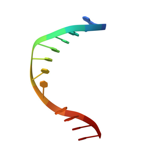
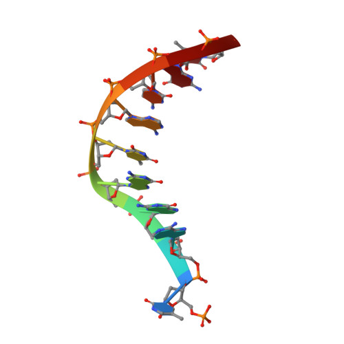
DNA (5'-D(*AP*CP*GP*TP*AP*CP*CP*TP*GP*GP*CP*T)-3')A [auth M]12N/A
Sequence Annotations
Expand
  • Reference Sequence

Find similar nucleic acids by:  Sequence   |   3D Structure  

Entity ID: 2
MoleculeChains LengthOrganismImage
DNA (5'-D(*AP*GP*C)-3')B [auth N]3N/A
Sequence Annotations
Expand
  • Reference Sequence

Find similar nucleic acids by:  Sequence   |   3D Structure  

Entity ID: 3
MoleculeChains LengthOrganismImage
DNA (5'-D(P*TP*AP*GP*GP*TP*AP*CP*GP*T)-3')C [auth O]9N/A
Sequence Annotations
Expand
  • Reference Sequence
Experimental Data & Validation

Experimental Data

  • Method: X-RAY DIFFRACTION
  • Resolution: 2.30 Å
  • R-Value Free: 0.230 
  • R-Value Work: 0.204 
  • R-Value Observed: 0.204 
  • Space Group: P 65
Unit Cell:
Length ( Å )Angle ( ˚ )
a = 63α = 90
b = 63β = 90
c = 152.5γ = 120
Software Package:
Software NamePurpose
AMoREphasing
CNSrefinement
DENZOdata reduction
SCALEPACKdata scaling

Structure Validation

View Full Validation Report


Entry History 

Deposition Data

Revision History  (Full details and data files)

  • Version 1.0: 1999-12-12
    Type: Initial release
  • Version 1.1: 2008-04-27
    Changes: Version format compliance
  • Version 1.2: 2011-07-13
    Changes: Version format compliance
  • Version 1.3: 2023-08-09
    Changes: Data collection, Database references, Derived calculations, Refinement description