1CG5

DEOXY FORM HEMOGLOBIN FROM DASYATIS AKAJEI


Experimental Data Snapshot

  • Method: X-RAY DIFFRACTION
  • Resolution: 1.60 Å
  • R-Value Free: 0.232 
  • R-Value Work: 0.191 

wwPDB Validation   3D Report Full Report


Ligand Structure Quality Assessment 


This is version 1.3 of the entry. See complete history


Literature

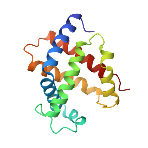
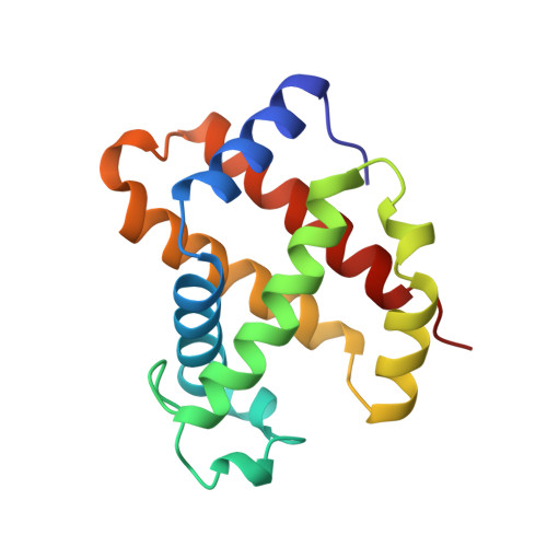
Structures of the deoxy and CO forms of haemoglobin from Dasyatis akajei, a cartilaginous fish.

Chong, K.T.Miyazaki, G.Morimoto, H.Oda, Y.Park, S.Y.

(1999) Acta Crystallogr D Biol Crystallogr 55: 1291-1300

  • DOI: https://doi.org/10.1107/s0907444999005934
  • Primary Citation of Related Structures:  
    1CG5, 1CG8

  • PubMed Abstract: 

    The three-dimensional structures of the deoxy- and carbonmonoxyhaemoglobin (Hb) from Dasyatis akajei, a stingray, have been determined at 1.6 and 1.9 A resolution, respectively. This is one of the most distantly related vertebrate Hbs to human HbA. Both structures resemble the respective forms of HbA, indicating that the alpha2beta2-type tetramer and the mode of the quaternary structure change are common to Hbs of jawed vertebrates. Larger deviations between D. akajei Hb and human HbA are observed in various parts of the molecule, even in the E and F helices. Significant mutations and/or conformational changes are also observed around the haems, in the C-terminal region of the beta subunit, in the alpha1beta2 interface and in the organic phosphate-binding site of HbA. Despite these structural differences, the oxygen affinity, haem-haem interaction, Bohr effect and organic phosphate effect of D. akajei Hb are all only moderately reduced. Compared with human HbA, the overall r.m.s. deviation of main-chain atoms in the helical regions of bony fish Hbs is smaller than that of D. akajei Hb.


  • Organizational Affiliation

    Division of Biophysical Engineering, Graduate School of Engineering Science, Osaka University, Toyonaka, Osaka 560-8531, Japan.


Macromolecules
Find similar proteins by:  (by identity cutoff)  |  3D Structure
Entity ID: 1
MoleculeChains Sequence LengthOrganismDetailsImage
PROTEIN (HEMOGLOBIN)141Hemitrygon akajeiMutation(s): 0 
UniProt
Find proteins for P56691 (Hemitrygon akajei)
Explore P56691 
Go to UniProtKB:  P56691
Entity Groups  
Sequence Clusters30% Identity50% Identity70% Identity90% Identity95% Identity100% Identity
UniProt GroupP56691
Sequence Annotations
Expand
  • Reference Sequence
Find similar proteins by:  (by identity cutoff)  |  3D Structure
Entity ID: 2
MoleculeChains Sequence LengthOrganismDetailsImage
PROTEIN (HEMOGLOBIN)141Hemitrygon akajeiMutation(s): 0 
UniProt
Find proteins for P56692 (Hemitrygon akajei)
Explore P56692 
Go to UniProtKB:  P56692
Entity Groups  
Sequence Clusters30% Identity50% Identity70% Identity90% Identity95% Identity100% Identity
UniProt GroupP56692
Sequence Annotations
Expand
  • Reference Sequence
Small Molecules
Ligands 1 Unique
IDChains Name / Formula / InChI Key2D Diagram3D Interactions
HEM
Query on HEM

Download Ideal Coordinates CCD File 
C [auth A],
D [auth B]
PROTOPORPHYRIN IX CONTAINING FE
C34 H32 Fe N4 O4
KABFMIBPWCXCRK-RGGAHWMASA-L
Experimental Data & Validation

Experimental Data

  • Method: X-RAY DIFFRACTION
  • Resolution: 1.60 Å
  • R-Value Free: 0.232 
  • R-Value Work: 0.191 
  • Space Group: P 31 1 2
Unit Cell:
Length ( Å )Angle ( ˚ )
a = 77.76α = 90
b = 77.76β = 90
c = 99.32γ = 120
Software Package:
Software NamePurpose
X-PLORmodel building
X-PLORrefinement
DENZOdata reduction
SCALEPACKdata scaling
X-PLORphasing

Structure Validation

View Full Validation Report


Ligand Structure Quality Assessment 


Entry History 

Deposition Data

Revision History  (Full details and data files)

  • Version 1.0: 1999-04-01
    Type: Initial release
  • Version 1.1: 2008-04-26
    Changes: Version format compliance
  • Version 1.2: 2011-07-13
    Changes: Version format compliance
  • Version 1.3: 2023-08-09
    Changes: Data collection, Database references, Derived calculations, Refinement description