1CD3 | pdb_00001cd3

PROCAPSID OF BACTERIOPHAGE PHIX174


Experimental Data Snapshot

  • Method: X-RAY DIFFRACTION
  • Resolution: 3.50 Å
  • R-Value Work: 
    0.275 (Depositor), 0.483 (DCC) 

Starting Model: experimental
View more details

wwPDB Validation 3D Report Full Report

Validation slider image for 1CD3

This is version 1.6 of the entry. See complete history

Literature

The role of scaffolding proteins in the assembly of the small, single-stranded DNA virus phiX174.

Dokland, T.Bernal, R.A.Burch, A.Pletnev, S.Fane, B.A.Rossmann, M.G.

(1999) J Mol Biology 288: 595-608

  • DOI: https://doi.org/10.1006/jmbi.1999.2699
  • Primary Citation Related Structures: 
    1CD3

  • PubMed Abstract: 

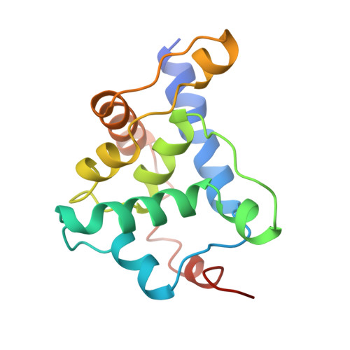
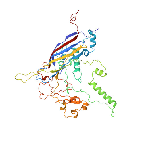
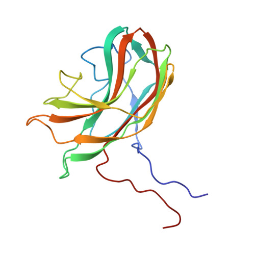
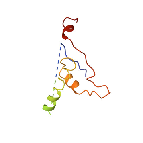
    An empty precursor particle called the procapsid is formed during assembly of the single-stranded DNA bacteriophage phiX174. Assembly of the phiX174 procapsid requires the presence of the two scaffolding proteins, D and B, which are structural components of the procapsid, but are not found in the mature virion. The X-ray crystallographic structure of a "closed" procapsid particle has been determined to 3.5 A resolution. This structure has an external scaffold made from 240 copies of protein D, 60 copies of the internally located B protein, and contains 60 copies of each of the viral structural proteins F and G, which comprise the shell and the 5-fold spikes, respectively. The F capsid protein has a similar conformation to that seen in the mature virion, and differs from the previously determined 25 A resolution electron microscopic reconstruction of the "open" procapsid, in which the F protein has a different conformation. The D scaffolding protein has a predominantly alpha-helical fold and displays remarkable conformational variability. We report here an improved and refined structure of the closed procapsid and describe in some detail the differences between the four independent D scaffolding proteins per icosahedral asymmetric unit, as well as their interaction with the F capsid protein. We re-analyze and correct the comparison of the closed procapsid with the previously determined cryo-electron microscopic image reconstruction of the open procapsid and discuss the major structural rearrangements that must occur during assembly. A model is proposed in which the D proteins direct the assembly process by sequential binding and conformational switching.


  • Organizational Affiliation
    • Department of Biological Sciences, Purdue University, West Lafayette, IN, 47907-1392, USA.

Macromolecule Content 

  • Total Structure Weight: 149.15 kDa 
  • Atom Count: 9,851 
  • Modeled Residue Count: 1,233 
  • Deposited Residue Count: 1,329 
  • Unique protein chains: 4

Macromolecules

Find similar proteins by:|  3D Structure
Entity ID: 1
MoleculeChains  Sequence LengthOrganismDetailsImage
PROTEIN (SCAFFOLDING PROTEIN GPD)A [auth 1],
B [auth 2],
C [auth 3],
D [auth 4]
152Sinsheimervirus phiX174Mutation(s): 0 
UniProt
Find proteins for P69486 (Enterobacteria phage phiX174)
Explore P69486 
Go to UniProtKB:  P69486
Entity Groups
Sequence Clusters30% Identity50% Identity70% Identity90% Identity95% Identity100% Identity
UniProt GroupP69486
Sequence Annotations
Expand
Reference Sequence
Find similar proteins by:|  3D Structure
Entity ID: 2
MoleculeChains  Sequence LengthOrganismDetailsImage
PROTEIN (CAPSID PROTEIN GPF)E [auth F]426Sinsheimervirus phiX174Mutation(s): 0 
UniProt
Find proteins for P03641 (Enterobacteria phage phiX174)
Explore P03641 
Go to UniProtKB:  P03641
Entity Groups
Sequence Clusters30% Identity50% Identity70% Identity90% Identity95% Identity100% Identity
UniProt GroupP03641
Sequence Annotations
Expand
Reference Sequence
Find similar proteins by:|  3D Structure
Entity ID: 3
MoleculeChains  Sequence LengthOrganismDetailsImage
PROTEIN (SPIKE PROTEIN GPG)F [auth G]175Sinsheimervirus phiX174Mutation(s): 0 
UniProt
Find proteins for P03643 (Enterobacteria phage phiX174)
Explore P03643 
Go to UniProtKB:  P03643
Entity Groups
Sequence Clusters30% Identity50% Identity70% Identity90% Identity95% Identity100% Identity
UniProt GroupP03643
Sequence Annotations
Expand
Reference Sequence
Find similar proteins by:|  3D Structure
Entity ID: 4
MoleculeChains  Sequence LengthOrganismDetailsImage
PROTEIN (SCAFFOLDING PROTEIN GPB)G [auth B]120Sinsheimervirus phiX174Mutation(s): 0 
EC: 3.4
UniProt
Find proteins for P03633 (Enterobacteria phage phiX174)
Explore P03633 
Go to UniProtKB:  P03633
Entity Groups
Sequence Clusters30% Identity50% Identity70% Identity90% Identity95% Identity100% Identity
UniProt GroupP03633
Sequence Annotations
Expand
Reference Sequence

Experimental Data & Validation

Experimental Data

  • Method: X-RAY DIFFRACTION
  • Resolution: 3.50 Å
  • R-Value Work:  0.275 (Depositor), 0.483 (DCC) 
Space Group: I 21 3
Unit Cell:
Length ( Å )Angle ( ˚ )
a = 774α = 90
b = 774β = 90
c = 774γ = 90
Software Package:
Software NamePurpose
MGRmodel building
X-PLORrefinement
DENZOdata reduction
SnBphasing
MGRphasing

Structure Validation

View Full Validation Report


Entry History 

Deposition Data

Revision History  (Full details and data files)

  • Version 1.0: 1999-04-14
    Type: Initial release
  • Version 1.1: 2008-04-26
    Changes: Version format compliance
  • Version 1.2: 2011-07-13
    Changes: Version format compliance
  • Version 1.3: 2017-10-04
    Changes: Refinement description
  • Version 1.4: 2019-11-06
    Changes: Data collection, Database references
  • Version 1.5: 2023-12-27
    Changes: Advisory, Data collection, Database references, Derived calculations
  • Version 1.6: 2024-04-03
    Changes: Refinement description