Yeast d-amino Acid oxidase: structural basis of its catalytic properties
Pollegioni, L., Diederichs, K., Molla, G., Umhau, S., Welte, W., Ghisla, S., Pilone, M.S.(2002) J Mol Biology 324: 535-546
- PubMed: 12445787 Search on PubMed
- DOI: https://doi.org/10.1016/s0022-2836(02)01062-8
- Primary Citation Related Structures:
1C0I - PubMed Abstract:
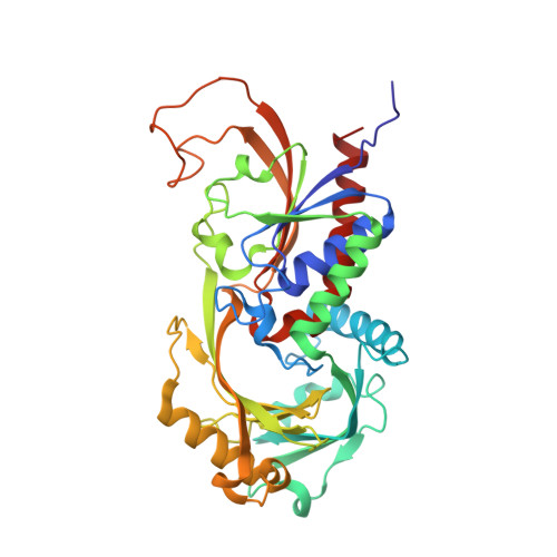
The 3D structure of the flavoprotein D-amino acid oxidase (DAAO) from the yeast Rhodotorula gracilis (RgDAAO) in complex with the competitive inhibitor anthranilate was solved (resolution 1.9A) and structural features relevant for the overall conformation and for catalytic activity are described. The FAD is bound in an elongated conformation in the core of the enzyme. Two anthranilate molecules are found within the active site cavity; one is located in a funnel forming the entrance, and the second is in contact with the flavin. The anchoring of the ligand carboxylate with Arg285 and Tyr223 is found for all complexes studied. However, while the active site group Tyr238-OH interacts with the carboxylate in the case of the substrate D-alanine, of D-CF(3)-alanine, or of L-lactate, in the anthranilate complex the phenol group rotates around the C2-C3 bond thus opening the entrance of the active site, and interacts there with the second bound anthranilate. This movement serves in channeling substrate to the bottom of the active site, the locus of chemical catalysis. The absence in RgDAAO of the "lid" covering the active site, as found in mammalian DAAO, is interpreted as being at the origin of the differences in kinetic mechanism between the two enzymes. This lid has been proposed to regulate product dissociation in the latter, while the side-chain of Tyr238 might exert a similar role in RgDAAO. The more open active site architecture of RgDAAO is the origin of its much broader substrate specificity. The RgDAAO enzyme forms a homodimer with C2 symmetry that is different from that reported for mammalian D-amino acid oxidase. This different mode of aggregation probably causes the differences in stability and tightness of FAD cofactor binding between the DAAOs from different sources.
- Department of Structural and Functional Biology, University of Insubria via J.H. Dunant, 3, 21100, Varese, Italy.
Organizational Affiliation: