1C08

CRYSTAL STRUCTURE OF HYHEL-10 FV-HEN LYSOZYME COMPLEX


Experimental Data Snapshot

  • Method: X-RAY DIFFRACTION
  • Resolution: 2.30 Å
  • R-Value Free: 0.175 
  • R-Value Work: 0.235 

wwPDB Validation   3D Report Full Report


This is version 1.3 of the entry. See complete history


Literature

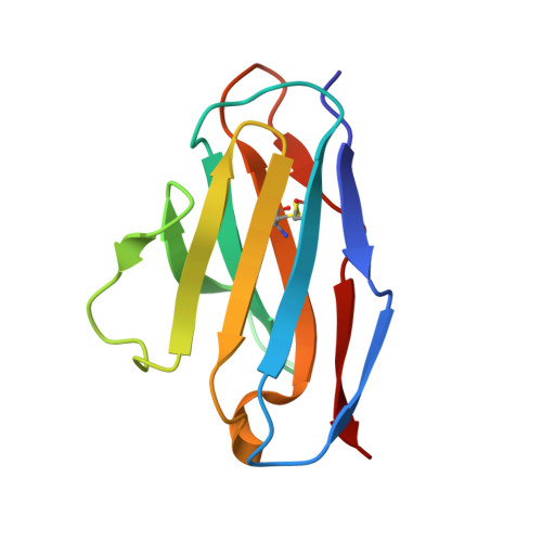
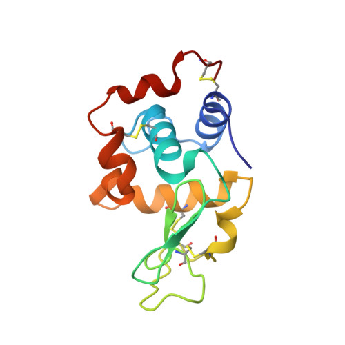
Crystal structure of anti-Hen egg white lysozyme antibody (HyHEL-10) Fv-antigen complex. Local structural changes in the protein antigen and water-mediated interactions of Fv-antigen and light chain-heavy chain interfaces.

Kondo, H.Shiroishi, M.Matsushima, M.Tsumoto, K.Kumagai, I.

(1999) J Biol Chem 274: 27623-27631

  • DOI: https://doi.org/10.1074/jbc.274.39.27623
  • Primary Citation of Related Structures:  
    1C08

  • PubMed Abstract: 

    In order to address the recognition mechanism of the fragments of antibody variable regions, termed Fv, toward their target antigen, an x-ray crystal structure of an anti-hen egg white lysozyme antibody (HyHEL-10) Fv fragment complexed with its cognate antigen, hen egg white lysozyme (HEL), was solved at 2.3 A. The overall structure of the complex is similar to that reported in a previous article dealing with the Fab fragment-HEL complex (PDB ID code,). However, the areas of Fv covered by HEL upon complex formation increased by about 100 A(2) in comparison with the Fab-HEL complex, and two local structural differences were observed in the heavy chain of the variable region (VH). In addition, small but significant local structural changes were observed in the antigen, HEL. The x-ray data permitted the identification of two water molecules between the VH and HEL and six water molecules retained in the interface between the antigen and the light chain complementarity determining regions (CDRs) 2 and 3 (CDR-L2 and CDR-L3). These water molecules bridge the antigen-antibody interface through hydrogen bond formation in the VL-HEL interface. Eleven water molecules were found to complete the imperfect VH-VL interface, suggesting that solvent molecules mediate the stabilization of interaction between variable regions. These results suggest that the unfavorable effect of deletion of constant regions on the antigen-antibody interaction is compensated by an increase in favorable interactions, including structural changes in the antigen-antibody interface and solvent-mediated hydrogen bond formation upon complex formation, which may lead to a minimum decreased affinity of the antibody Fv fragment toward its antigen.


  • Organizational Affiliation

    Department of Biomolecular Engineering, Graduate School of Engineering, Tohoku University, Aoba-yama 07, Sendai 980-8579, Japan.


Macromolecules
Find similar proteins by:  (by identity cutoff)  |  3D Structure
Entity ID: 1
MoleculeChains Sequence LengthOrganismDetailsImage
ANTI-HEN EGG WHITE LYSOZYME ANTIBODY (HYHEL-10)107Mus musculusMutation(s): 0 
UniProt
Find proteins for P01642 (Mus musculus)
Explore P01642 
Go to UniProtKB:  P01642
Entity Groups  
Sequence Clusters30% Identity50% Identity70% Identity90% Identity95% Identity100% Identity
UniProt GroupP01642
Sequence Annotations
Expand
  • Reference Sequence
Find similar proteins by:  (by identity cutoff)  |  3D Structure
Entity ID: 2
MoleculeChains Sequence LengthOrganismDetailsImage
ANTI-HEN EGG WHITE LYSOZYME ANTIBODY (HYHEL-10)114Mus musculusMutation(s): 0 
UniProt
Find proteins for P01823 (Mus musculus)
Explore P01823 
Go to UniProtKB:  P01823
Entity Groups  
Sequence Clusters30% Identity50% Identity70% Identity90% Identity95% Identity100% Identity
UniProt GroupP01823
Sequence Annotations
Expand
  • Reference Sequence
Find similar proteins by:  (by identity cutoff)  |  3D Structure
Entity ID: 3
MoleculeChains Sequence LengthOrganismDetailsImage
LYSOZYME129Gallus gallusMutation(s): 0 
UniProt
Find proteins for P00698 (Gallus gallus)
Explore P00698 
Go to UniProtKB:  P00698
Entity Groups  
Sequence Clusters30% Identity50% Identity70% Identity90% Identity95% Identity100% Identity
UniProt GroupP00698
Sequence Annotations
Expand
  • Reference Sequence
Experimental Data & Validation

Experimental Data

  • Method: X-RAY DIFFRACTION
  • Resolution: 2.30 Å
  • R-Value Free: 0.175 
  • R-Value Work: 0.235 
  • Space Group: P 41 21 2
Unit Cell:
Length ( Å )Angle ( ˚ )
a = 57.208α = 90
b = 57.208β = 90
c = 236.071γ = 90
Software Package:
Software NamePurpose
DENZOdata reduction
SCALAdata scaling
AMoREphasing
REFMACrefinement
CCP4data scaling

Structure Validation

View Full Validation Report


Entry History 

Deposition Data

Revision History  (Full details and data files)

  • Version 1.0: 2000-07-19
    Type: Initial release
  • Version 1.1: 2008-04-27
    Changes: Version format compliance
  • Version 1.2: 2011-07-13
    Changes: Version format compliance
  • Version 1.3: 2023-05-31
    Changes: Database references