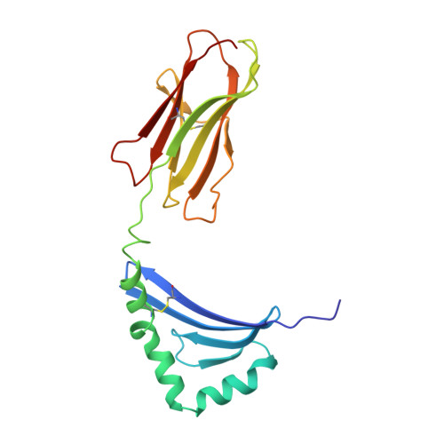
1BX2

CRYSTAL STRUCTURE OF HLA-DR2 (DRA*0101,DRB1*1501) COMPLEXED WITH A PEPTIDE FROM HUMAN MYELIN BASIC PROTEIN


Experimental Data Snapshot

  • Method: X-RAY DIFFRACTION
  • Resolution: 2.60 Å
  • R-Value Free: 0.266 
  • R-Value Work: 0.238 
  • R-Value Observed: 0.238 

Starting Model: experimental
View more details

wwPDB Validation   3D Report Full Report


This is version 1.4 of the entry. See complete history


Literature

Crystal structure of HLA-DR2 (DRA*0101, DRB1*1501) complexed with a peptide from human myelin basic protein.

Smith, K.J.Pyrdol, J.Gauthier, L.Wiley, D.C.Wucherpfennig, K.W.

(1998) J Exp Med 188: 1511-1520

  • DOI: https://doi.org/10.1084/jem.188.8.1511
  • Primary Citation of Related Structures:  
    1BX2

  • PubMed Abstract: 

    Susceptibility to multiple sclerosis is associated with the human histocompatibility leukocyte antigen (HLA)-DR2 (DRB1*1501) haplotype. The structure of HLA-DR2 was determined with a bound peptide from human myelin basic protein (MBP) that is immunodominant for human MBP-specific T cells. Residues of MBP peptide that are important for T cell receptor recognition are prominent, solvent exposed residues in the crystal structure. A distinguishing feature of the HLA-DR2 peptide binding site is a large, primarily hydrophobic P4 pocket that accommodates a phenylalanine of the MBP peptide. The necessary space for this aromatic side chain is created by an alanine at the polymorphic DRbeta 71 position. These features make the P4 pocket of HLA-DR2 distinct from DR molecules associated with other autoimmune diseases.


  • Organizational Affiliation

    Department of Molecular Medicine, Children's Hospital, Boston, Massachusetts 02115, USA. ksmith@rascal.med.harvard.edu


Macromolecules
Find similar proteins by:  (by identity cutoff)  |  3D Structure
Entity ID: 1
MoleculeChains Sequence LengthOrganismDetailsImage
PROTEIN (HLA-DR2)
A, D
180Homo sapiensMutation(s): 0 
UniProt & NIH Common Fund Data Resources
Find proteins for P01903 (Homo sapiens)
Explore P01903 
Go to UniProtKB:  P01903
PHAROS:  P01903
GTEx:  ENSG00000204287 
Entity Groups  
Sequence Clusters30% Identity50% Identity70% Identity90% Identity95% Identity100% Identity
UniProt GroupP01903
Glycosylation
Glycosylation Sites: 1Go to GlyGen: P01903-1
Sequence Annotations
Expand
  • Reference Sequence
Find similar proteins by:  (by identity cutoff)  |  3D Structure
Entity ID: 2
MoleculeChains Sequence LengthOrganismDetailsImage
PROTEIN (HLA-DR2)
B, E
191Homo sapiensMutation(s): 0 
UniProt & NIH Common Fund Data Resources
Find proteins for P01911 (Homo sapiens)
Explore P01911 
Go to UniProtKB:  P01911
PHAROS:  P01911
GTEx:  ENSG00000196126 
Entity Groups  
Sequence Clusters30% Identity50% Identity70% Identity90% Identity95% Identity100% Identity
UniProt GroupP01911
Sequence Annotations
Expand
  • Reference Sequence

Find similar proteins by:  Sequence   |   3D Structure  

Entity ID: 3
MoleculeChains Sequence LengthOrganismDetailsImage
PROTEIN (HLA-DR2)
C, F
15Homo sapiensMutation(s): 0 
UniProt & NIH Common Fund Data Resources
Find proteins for P02686 (Homo sapiens)
Explore P02686 
Go to UniProtKB:  P02686
PHAROS:  P02686
GTEx:  ENSG00000197971 
Entity Groups  
Sequence Clusters30% Identity50% Identity70% Identity90% Identity95% Identity100% Identity
UniProt GroupP02686
Sequence Annotations
Expand
  • Reference Sequence
Small Molecules
Ligands 1 Unique
IDChains Name / Formula / InChI Key2D Diagram3D Interactions
NAG
Query on NAG

Download Ideal Coordinates CCD File 
G [auth A],
H [auth D]
2-acetamido-2-deoxy-beta-D-glucopyranose
C8 H15 N O6
OVRNDRQMDRJTHS-FMDGEEDCSA-N
Experimental Data & Validation

Experimental Data

  • Method: X-RAY DIFFRACTION
  • Resolution: 2.60 Å
  • R-Value Free: 0.266 
  • R-Value Work: 0.238 
  • R-Value Observed: 0.238 
  • Space Group: P 43 21 2
Unit Cell:
Length ( Å )Angle ( ˚ )
a = 95.403α = 90
b = 95.403β = 90
c = 295.219γ = 90
Software Package:
Software NamePurpose
DENZOdata reduction
SCALEPACKdata scaling
AMoREphasing
X-PLORrefinement

Structure Validation

View Full Validation Report


Entry History 

Deposition Data

Revision History  (Full details and data files)

  • Version 1.0: 1998-10-21
    Type: Initial release
  • Version 1.1: 2008-04-26
    Changes: Version format compliance
  • Version 1.2: 2011-07-13
    Changes: Derived calculations, Version format compliance
  • Version 1.3: 2020-07-29
    Type: Remediation
    Reason: Carbohydrate remediation
    Changes: Data collection, Derived calculations, Structure summary
  • Version 1.4: 2023-08-09
    Changes: Data collection, Database references, Refinement description, Structure summary