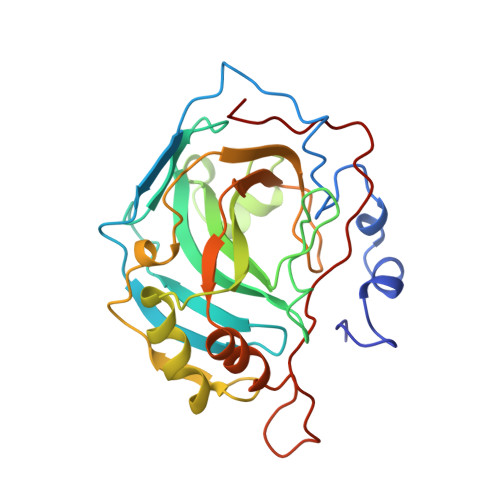
1BN1

CARBONIC ANHYDRASE II INHIBITOR


Experimental Data Snapshot

  • Method: X-RAY DIFFRACTION
  • Resolution: 2.10 Å
  • R-Value Work: 0.166 
  • R-Value Observed: 0.166 

Starting Model: experimental
View more details

wwPDB Validation   3D Report Full Report


This is version 1.4 of the entry. See complete history


Literature

Structural analysis of inhibitor binding to human carbonic anhydrase II.

Boriack-Sjodin, P.A.Zeitlin, S.Chen, H.H.Crenshaw, L.Gross, S.Dantanarayana, A.Delgado, P.May, J.A.Dean, T.Christianson, D.W.

(1998) Protein Sci 7: 2483-2489

  • DOI: https://doi.org/10.1002/pro.5560071201
  • Primary Citation of Related Structures:  
    1BN1, 1BN3, 1BN4, 1BNM, 1BNN, 1BNQ, 1BNT, 1BNU, 1BNV, 1BNW

  • PubMed Abstract: 

    X-ray crystal structures of carbonic anhydrase II (CAII) complexed with sulfonamide inhibitors illuminate the structural determinants of high affinity binding in the nanomolar regime. The primary binding interaction is the coordination of a primary sulfonamide group to the active site zinc ion. Secondary interactions fine-tune tight binding in regions of the active site cavity >5 A away from zinc, and this work highlights three such features: (1) advantageous conformational restraints of a bicyclic thienothiazene-6-sulfonamide-1,1-dioxide inhibitor skeleton in comparison with a monocyclic 2,5-thiophenedisulfonamide skeleton; (2) optimal substituents attached to a secondary sulfonamide group targeted to interact with hydrophobic patches defined by Phe131, Leu198, and Pro202; and (3) optimal stereochemistry and configuration at the C-4 position of bicyclic thienothiazene-6-sulfonamides; the C-4 substituent can interact with His64, the catalytic proton shuttle. Structure-activity relationships rationalize affinity trends observed during the development of brinzolamide (Azopt), the newest carbonic anhydrase inhibitor approved for the treatment of glaucoma.


  • Organizational Affiliation

    Department of Chemistry, University of Pennsylvania, Philadelphia 19104-6323, USA.


Macromolecules
Find similar proteins by:  (by identity cutoff)  |  3D Structure
Entity ID: 1
MoleculeChains Sequence LengthOrganismDetailsImage
CARBONIC ANHYDRASE259Homo sapiensMutation(s): 0 
EC: 4.2.1.1
UniProt & NIH Common Fund Data Resources
Find proteins for P00918 (Homo sapiens)
Explore P00918 
Go to UniProtKB:  P00918
PHAROS:  P00918
GTEx:  ENSG00000104267 
Entity Groups  
Sequence Clusters30% Identity50% Identity70% Identity90% Identity95% Identity100% Identity
UniProt GroupP00918
Sequence Annotations
Expand
  • Reference Sequence
Small Molecules
Ligands 3 Unique
IDChains Name / Formula / InChI Key2D Diagram3D Interactions
AL5
Query on AL5

Download Ideal Coordinates CCD File 
D [auth A]THIOPHENE-2,5-DISULFONIC ACID 2-AMIDE-5-(4-METHYL-BENZYLAMIDE)
C12 H14 N2 O4 S3
GUNRMHMEAWSZQE-UHFFFAOYSA-N
HG
Query on HG

Download Ideal Coordinates CCD File 
B [auth A]MERCURY (II) ION
Hg
BQPIGGFYSBELGY-UHFFFAOYSA-N
ZN
Query on ZN

Download Ideal Coordinates CCD File 
C [auth A]ZINC ION
Zn
PTFCDOFLOPIGGS-UHFFFAOYSA-N
Binding Affinity Annotations 
IDSourceBinding Affinity
AL5 PDBBind:  1BN1 Kd: 0.46 (nM) from 1 assay(s)
Experimental Data & Validation

Experimental Data

  • Method: X-RAY DIFFRACTION
  • Resolution: 2.10 Å
  • R-Value Work: 0.166 
  • R-Value Observed: 0.166 
  • Space Group: P 1 21 1
Unit Cell:
Length ( Å )Angle ( ˚ )
a = 42.7α = 90
b = 41.7β = 104.6
c = 73γ = 90
Software Package:
Software NamePurpose
X-PLORmodel building
X-PLORrefinement
MOSFLMdata reduction
CCP4data scaling
X-PLORphasing

Structure Validation

View Full Validation Report


Entry History 

Deposition Data

Revision History  (Full details and data files)

  • Version 1.0: 1999-05-18
    Type: Initial release
  • Version 1.1: 2008-03-03
    Changes: Version format compliance
  • Version 1.2: 2011-07-13
    Changes: Version format compliance
  • Version 1.3: 2023-08-09
    Changes: Database references, Derived calculations, Other, Refinement description
  • Version 1.4: 2024-05-22
    Changes: Data collection