1BM3 | pdb_00001bm3

IMMUNOGLOBULIN OPG2 FAB-PEPTIDE COMPLEX


Experimental Data Snapshot

  • Method: X-RAY DIFFRACTION
  • Resolution: 2.00 Å
  • R-Value Work: 
    0.152 (Depositor) 

Starting Model: experimental
View more details

wwPDB Validation 3D Report Full Report

Validation slider image for 1BM3

This is version 1.6 of the entry. See complete history

Literature

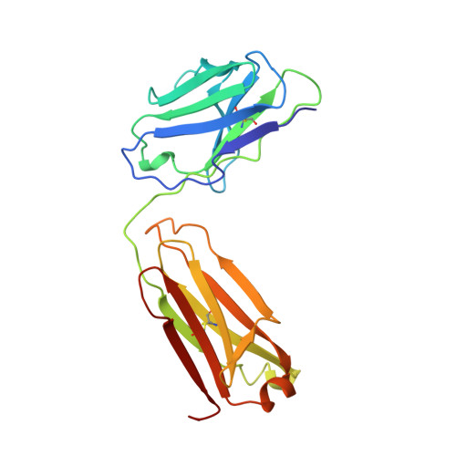
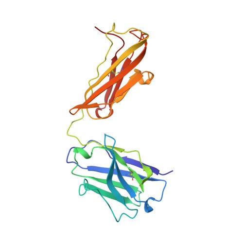
Conformational change in an anti-integrin antibody: structure of OPG2 Fab bound to a beta 3 peptide.

Kodandapani, R.Veerapandian, L.Ni, C.Z.Chiou, C.K.Whittal, R.M.Kunicki, T.J.Ely, K.R.

(1998) Biochem Biophys Res Commun 251: 61-66

  • DOI: https://doi.org/10.1006/bbrc.1998.9380
  • Primary Citation Related Structures: 
    1BM3

  • PubMed Abstract: 

    Antibodies are important tools to explore receptor-ligand interactions. The anti-integrin antibody OPG2 binds in an RGD-related manner to the alphaIIb beta3 integrin as a molecular mimic of fibrinogen. The Fab fragment from OPG2 was cocrystallized with a peptide from the beta3 subunit of the integrin representing a site that binds RGD. The crystal structure of the complex was determined at 2.2-A resolution and compared with the unbound Fab. On binding the integrin peptide there were conformational changes in CDR3 of the heavy chain. Also, a significant shift across the intermolecular interface between the CH1-CL domains was observed so that the angle of rotation relating the two domains was reduced by 15 degrees. This unusual conformational adjustment represents the first example of ligand-induced conformational changes in the carboxyl domains of a Fab fragment.


  • Organizational Affiliation
    • Cancer Research Center, The Burnham Institute, La Jolla, California, 92037, USA.

Macromolecule Content 

  • Total Structure Weight: 48.06 kDa 
  • Atom Count: 3,589 
  • Modeled Residue Count: 430 
  • Deposited Residue Count: 441 
  • Unique protein chains: 2

Macromolecules

Find similar proteins by:|  3D Structure
Entity ID: 1
MoleculeChains  Sequence LengthOrganismDetailsImage
IMMUNOGLOBULIN OPG2 FAB, CONSTANT DOMAINA [auth L]214Mus musculusMutation(s): 0 
Find similar proteins by:|  3D Structure
Entity ID: 2
MoleculeChains  Sequence LengthOrganismDetailsImage
IMMUNOGLOBULIN OPG2 FAB, VARIABLE DOMAINB [auth H]227Mus musculusMutation(s): 0 
UniProt
Find proteins for P01868 (Mus musculus)
Explore P01868 
Go to UniProtKB:  P01868
Entity Groups
Sequence Clusters30% Identity50% Identity70% Identity90% Identity95% Identity100% Identity
UniProt GroupP01868
Sequence Annotations
Expand
Reference Sequence

Experimental Data & Validation

Experimental Data

  • Method: X-RAY DIFFRACTION
  • Resolution: 2.00 Å
  • R-Value Work:  0.152 (Depositor) 
Space Group: A 1 2 1
Unit Cell:
Length ( Å )Angle ( ˚ )
a = 74.11α = 90
b = 90.05β = 115.3
c = 79.11γ = 90
Software Package:
Software NamePurpose
UCSD-systemdata collection
NILSENdata reduction
X-PLORrefinement
UCSD-systemdata reduction
X-GENdata scaling
NILSENdata scaling

Structure Validation

View Full Validation Report


Entry History 

Deposition Data

Revision History  (Full details and data files)

  • Version 1.0: 1999-04-20
    Type: Initial release
  • Version 1.1: 2008-04-26
    Changes: Version format compliance
  • Version 1.2: 2011-07-13
    Changes: Version format compliance
  • Version 1.3: 2017-10-04
    Changes: Refinement description
  • Version 1.4: 2019-11-27
    Changes: Database references
  • Version 1.5: 2023-08-09
    Changes: Data collection, Database references, Derived calculations, Refinement description
  • Version 1.6: 2024-10-30
    Changes: Structure summary