1B5X

Contribution of hydrogen bonds to the conformational stability of human lysozyme: calorimetry and x-ray analysis of six ser->ala mutants


Experimental Data Snapshot

  • Method: X-RAY DIFFRACTION
  • Resolution: 2.00 Å
  • R-Value Work: 0.152 
  • R-Value Observed: 0.152 

wwPDB Validation   3D Report Full Report


This is version 1.2 of the entry. See complete history


Literature

Contribution of hydrogen bonds to the conformational stability of human lysozyme: calorimetry and X-ray analysis of six Ser --> Ala mutants.

Takano, K.Yamagata, Y.Kubota, M.Funahashi, J.Fujii, S.Yutani, K.

(1999) Biochemistry 38: 6623-6629

  • DOI: https://doi.org/10.1021/bi9901228
  • Primary Citation of Related Structures:  
    1B5U, 1B5V, 1B5W, 1B5X, 1B5Y, 1B5Z

  • PubMed Abstract: 

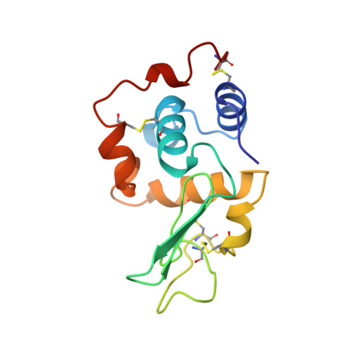
    To further examine the contribution of hydrogen bonds to the conformational stability of the human lysozyme, six Ser to Ala mutants were constructed. The thermodynamic parameters for denaturation of these six Ser mutant proteins were investigated by differential scanning calorimetry (DSC), and the crystal structures were determined by X-ray analysis. The denaturation Gibbs energy (DeltaG) of the Ser mutant proteins was changed from 2.0 to -5.7 kJ/mol, compared to that of the wild-type protein. With an analysis in which some factors that affected the stability due to mutation were considered, the contribution of hydrogen bonds to the stability (Delta DeltaGHB) was extracted on the basis of the structures of the mutant proteins. The results showed that hydrogen bonds between protein atoms and between a protein atom and a water bound with the protein molecule favorably contribute to the protein stability. The net contribution of one intramolecular hydrogen bond to protein stability (DeltaGHB) was 8.9 +/- 2.6 kJ/mol on average. However, the contribution to the protein stability of hydrogen bonds between a protein atom and a bound water molecule was smaller than that for a bond between protein atoms.


  • Organizational Affiliation

    Institute for Protein Research, Osaka University, Japan.


Macromolecules
Find similar proteins by:  (by identity cutoff)  |  3D Structure
Entity ID: 1
MoleculeChains Sequence LengthOrganismDetailsImage
PROTEIN (LYSOZYME)130Homo sapiensMutation(s): 0 
EC: 3.2.1.17
UniProt & NIH Common Fund Data Resources
Find proteins for P61626 (Homo sapiens)
Explore P61626 
Go to UniProtKB:  P61626
PHAROS:  P61626
GTEx:  ENSG00000090382 
Entity Groups  
Sequence Clusters30% Identity50% Identity70% Identity90% Identity95% Identity100% Identity
UniProt GroupP61626
Sequence Annotations
Expand
  • Reference Sequence
Small Molecules
Ligands 1 Unique
IDChains Name / Formula / InChI Key2D Diagram3D Interactions
NA
Query on NA

Download Ideal Coordinates CCD File 
B [auth A]SODIUM ION
Na
FKNQFGJONOIPTF-UHFFFAOYSA-N
Experimental Data & Validation

Experimental Data

  • Method: X-RAY DIFFRACTION
  • Resolution: 2.00 Å
  • R-Value Work: 0.152 
  • R-Value Observed: 0.152 
  • Space Group: P 21 21 21
Unit Cell:
Length ( Å )Angle ( ˚ )
a = 56.36α = 90
b = 62.62β = 90
c = 32.55γ = 90
Software Package:
Software NamePurpose
X-PLORmodel building
X-PLORrefinement
X-PLORphasing

Structure Validation

View Full Validation Report


Entry History 

Deposition Data

Revision History  (Full details and data files)

  • Version 1.0: 1999-01-20
    Type: Initial release
  • Version 1.1: 2008-04-27
    Changes: Version format compliance
  • Version 1.2: 2011-07-13
    Changes: Version format compliance