1AII | pdb_00001aii

ANNEXIN III


Experimental Data Snapshot

  • Method: X-RAY DIFFRACTION
  • Resolution: 1.95 Å
  • R-Value Free: 
    0.237 (Depositor), 0.171 (DCC) 
  • R-Value Work: 
    0.193 (Depositor), 0.166 (DCC) 
  • R-Value Observed: 
    0.196 (Depositor) 

Starting Model: experimental
View more details

wwPDB Validation 3D Report Full Report

Validation slider image for 1AII

This is version 2.0 of the entry. See complete history

Literature

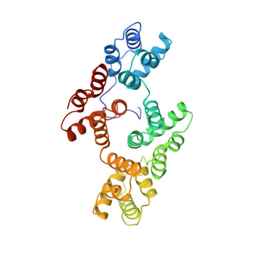
Can enzymatic activity, or otherwise, be inferred from structural studies of annexin III?

Perron, B.Lewit-Bentley, A.Geny, B.Russo-Marie, F.

(1997) J Biological Chem 272: 11321-11326

  • DOI: https://doi.org/10.1074/jbc.272.17.11321
  • Primary Citation Related Structures: 
    1AII

  • PubMed Abstract: 

    Annexin III, a putative inositol (1,2)-phosphohydrolase, was co-crystallized with inositol 2-phosphate, the inhibitor of the reaction, and its structure was solved to 1.95 A resolution. No enzyme active site was observed in the structure. Assays for enzymatic activity were also negative. Search for annexin III-inositol phosphate interactions using the BIAcoreTM system revealed an affinity for inositol cyclic (1,2)-phosphate, suggesting annexin III may sequester the molecule in the cell. The BIAcoreTM system used with different phospholipids showed that annexin III displays specificity for phosphatidylethanolamine, but not for phosphatidylinositols. Interestingly, a molecule of ethanolamine was found bound to the protein in the crystal structure. Coupled with the fact that this is a particularly abundant phospholipid in granules specific to neutrophils, cells where annexin III is highly expressed, our finding could be pointing to a physiological role of annexin III.


  • Organizational Affiliation
    • Institut Cochin de Génétique Moléculaire, U332 INSERM, 22 rue Méchain, 75014 Paris, France.

Macromolecule Content 

  • Total Structure Weight: 36.78 kDa 
  • Atom Count: 2,802 
  • Modeled Residue Count: 322 
  • Deposited Residue Count: 323 
  • Unique protein chains: 1

Macromolecules

Find similar proteins by:|  3D Structure
Entity ID: 1
MoleculeChains  Sequence LengthOrganismDetailsImage
ANNEXIN III323Homo sapiensMutation(s): 0 
UniProt & NIH Common Fund Data Resources
Find proteins for P12429 (Homo sapiens)
Explore P12429 
Go to UniProtKB:  P12429
PHAROS:  P12429
GTEx:  ENSG00000138772 
Entity Groups
Sequence Clusters30% Identity50% Identity70% Identity90% Identity95% Identity100% Identity
UniProt GroupP12429
Sequence Annotations
Expand
Reference Sequence

Small Molecules

Ligands 3 Unique
IDChains Name / Formula / InChI Key2D Diagram3D Interactions
SO4

Query on SO4


Download:Ideal Coordinates CCD File
G [auth A]SULFATE ION
O4 S
QAOWNCQODCNURD-UHFFFAOYSA-L
ETA

Query on ETA


Download:Ideal Coordinates CCD File
H [auth A]ETHANOLAMINE
C2 H7 N O
HZAXFHJVJLSVMW-UHFFFAOYSA-N
CA

Query on CA


Download:Ideal Coordinates CCD File
B [auth A],
C [auth A],
D [auth A],
E [auth A],
F [auth A]
CALCIUM ION
Ca
BHPQYMZQTOCNFJ-UHFFFAOYSA-N

Experimental Data & Validation

Experimental Data

  • Method: X-RAY DIFFRACTION
  • Resolution: 1.95 Å
  • R-Value Free:  0.237 (Depositor), 0.171 (DCC) 
  • R-Value Work:  0.193 (Depositor), 0.166 (DCC) 
  • R-Value Observed: 0.196 (Depositor) 
Space Group: P 1 21 1
Unit Cell:
Length ( Å )Angle ( ˚ )
a = 42.29α = 90
b = 68.72β = 94.27
c = 50.59γ = 90
Software Package:
Software NamePurpose
AMoREphasing
PROLSQrefinement
MOSFLMdata reduction
CCP4data reduction
CCP4data scaling

Structure Validation

View Full Validation Report


Entry History 

Deposition Data

Revision History  (Full details and data files)

  • Version 1.0: 1997-03-12
    Type: Initial release
  • Version 1.1: 2008-03-24
    Changes: Version format compliance
  • Version 1.2: 2011-07-13
    Changes: Version format compliance
  • Version 1.3: 2023-08-02
    Changes: Database references, Derived calculations, Refinement description
  • Version 2.0: 2023-11-15
    Changes: Atomic model, Data collection