12CA

ALTERING THE MOUTH OF A HYDROPHOBIC POCKET. STRUCTURE AND KINETICS OF HUMAN CARBONIC ANHYDRASE II MUTANTS AT RESIDUE VAL-121


Experimental Data Snapshot

  • Method: X-RAY DIFFRACTION
  • Resolution: 2.40 Å
  • R-Value Observed: 0.170 

wwPDB Validation   3D Report Full Report


This is version 1.4 of the entry. See complete history


Literature

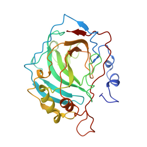
Altering the mouth of a hydrophobic pocket. Structure and kinetics of human carbonic anhydrase II mutants at residue Val-121.

Nair, S.K.Calderone, T.L.Christianson, D.W.Fierke, C.A.

(1991) J Biol Chem 266: 17320-17325

  • Primary Citation of Related Structures:  
    12CA

  • PubMed Abstract: 

    Eleven amino acid substitutions at Val-121 of human carbonic anhydrase II including Gly, Ala, Ser, Leu, Ile, Lys, and Arg, were constructed by site-directed mutagenesis. This residue is at the mouth of the hydrophobic pocket in the enzyme active site. The CO2 hydrase activity and the p-nitrophenyl esterase activity of these CAII variants correlate with the hydrophobicity of the residue, suggesting that the hydrophobic character of this residue is important for catalysis. The effects of these mutations on the steady-state kinetics for CO2 hydration occur mainly in kcat/Km and Km, consistent with involvement of this residue in CO2 association. The Val-121----Ala mutant, which exhibits about one-third normal CO2 hydrase activity, has been studied by x-ray crystallographic methods. No significant changes in the mutant enzyme conformation are evident relative to the wild-type enzyme. Since Val-121 is at the mouth of the hydrophobic pocket, its substitution by the methyl side chain of alanine makes the pocket mouth significantly wider than that of the wild-type enzyme. Hence, although a moderately wide (and deep) pocket is important for substrate association, a wider mouth to this pocket does not seriously compromise the catalytic approach of CO2 toward nucleophilic zinc-bound hydroxide.


  • Organizational Affiliation

    Department of Chemistry, University of Pennsylvania, Philadelphia 19104-6323.


Macromolecules
Find similar proteins by:  (by identity cutoff)  |  3D Structure
Entity ID: 1
MoleculeChains Sequence LengthOrganismDetailsImage
CARBONIC ANHYDRASE II260Homo sapiensMutation(s): 0 
EC: 4.2.1.1
UniProt & NIH Common Fund Data Resources
Find proteins for P00918 (Homo sapiens)
Explore P00918 
Go to UniProtKB:  P00918
PHAROS:  P00918
GTEx:  ENSG00000104267 
Entity Groups  
Sequence Clusters30% Identity50% Identity70% Identity90% Identity95% Identity100% Identity
UniProt GroupP00918
Sequence Annotations
Expand
  • Reference Sequence
Small Molecules
Ligands 2 Unique
IDChains Name / Formula / InChI Key2D Diagram3D Interactions
HG
Query on HG

Download Ideal Coordinates CCD File 
C [auth A]MERCURY (II) ION
Hg
BQPIGGFYSBELGY-UHFFFAOYSA-N
ZN
Query on ZN

Download Ideal Coordinates CCD File 
B [auth A]ZINC ION
Zn
PTFCDOFLOPIGGS-UHFFFAOYSA-N
Experimental Data & Validation

Experimental Data

  • Method: X-RAY DIFFRACTION
  • Resolution: 2.40 Å
  • R-Value Observed: 0.170 
  • Space Group: P 1 21 1
Unit Cell:
Length ( Å )Angle ( ˚ )
a = 42.7α = 90
b = 41.7β = 104.6
c = 73γ = 90
Software Package:
Software NamePurpose
PROLSQrefinement

Structure Validation

View Full Validation Report


Entry History 

Deposition Data

Revision History  (Full details and data files)

  • Version 1.0: 1992-10-15
    Type: Initial release
  • Version 1.1: 2008-03-24
    Changes: Version format compliance
  • Version 1.2: 2011-07-13
    Changes: Version format compliance
  • Version 1.3: 2017-11-29
    Changes: Derived calculations, Other
  • Version 1.4: 2024-02-07
    Changes: Data collection, Database references, Derived calculations