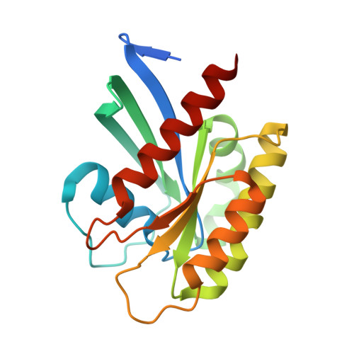
8VM2

Crystal Structure of NRAS Q61K bound to GTP


Experimental Data Snapshot

  • Method: X-RAY DIFFRACTION
  • Resolution: 1.74 Å
  • R-Value Free: 0.203 
  • R-Value Work: 0.172 
  • R-Value Observed: 0.173 

wwPDB Validation   3D Report Full Report


Ligand Structure Quality Assessment 


This is version 1.0 of the entry. See complete history


Literature

Crystal structure of NRAS Q61K with a ligand-induced pocket near switch II.

Gebregiworgis, T.Chan, J.Y.Kuntz, D.A.Prive, G.G.Marshall, C.B.Ikura, M.

(2024) Eur J Cell Biol 103: 151414-151414

  • DOI: https://doi.org/10.1016/j.ejcb.2024.151414
  • Primary Citation of Related Structures:  
    8VM2

  • PubMed Abstract: 

    The RAS isoforms (KRAS, HRAS and NRAS) have distinct cancer type-specific profiles. NRAS mutations are the second most prevalent RAS mutations in skin and hematological malignancies. Although RAS proteins were considered undruggable for decades, isoform and mutation-specific investigations have produced successful RAS inhibitors that are either specific to certain mutants, isoforms (pan-KRAS) or target all RAS proteins (pan-RAS). While extensive structural and biochemical investigations have focused mainly on K- and H-RAS mutations, NRAS mutations have received less attention, and the most prevalent NRAS mutations in human cancers, Q61K and Q61R, are rare in K- and H-RAS. This manuscript presents a crystal structure of the NRAS Q61K mutant in the GTP-bound form. Our structure reveals a previously unseen pocket near switch II induced by the binding of a ligand to the active form of the protein. This observation reveals a binding site that can potentially be exploited for development of inhibitors against mutant NRAS. Furthermore, the well-resolved catalytic site of this GTPase bound to native GTP provides insight into the stalled GTP hydrolysis observed for NRAS-Q61K.


  • Organizational Affiliation

    Princess Margaret Cancer Centre, University Health Network, Toronto, Ontario M5G 1L7, Canada; Department of Biochemistry, Schulich School of Medicine and Dentistry, Western University, London, Ontario N6A 5C1, Canada; Department of Oncology, Schulich School of Medicine and Dentistry, Western University, London, Ontario N6A 5W9, Canada. Electronic address: tgebregi@uwo.ca.


Macromolecules
Find similar proteins by:  (by identity cutoff)  |  3D Structure
Entity ID: 1
MoleculeChains Sequence LengthOrganismDetailsImage
GTPase NRas
A, B, C
190Homo sapiensMutation(s): 1 
Gene Names: NRASHRAS1
EC: 3.6.5.2
UniProt & NIH Common Fund Data Resources
Find proteins for P01111 (Homo sapiens)
Explore P01111 
Go to UniProtKB:  P01111
PHAROS:  P01111
GTEx:  ENSG00000213281 
Entity Groups  
Sequence Clusters30% Identity50% Identity70% Identity90% Identity95% Identity100% Identity
UniProt GroupP01111
Sequence Annotations
Expand
  • Reference Sequence
Small Molecules
Ligands 3 Unique
IDChains Name / Formula / InChI Key2D Diagram3D Interactions
GTP (Subject of Investigation/LOI)
Query on GTP

Download Ideal Coordinates CCD File 
D [auth A],
F [auth A],
H [auth B],
J [auth B],
K [auth C]
GUANOSINE-5'-TRIPHOSPHATE
C10 H16 N5 O14 P3
XKMLYUALXHKNFT-UUOKFMHZSA-N
MG
Query on MG

Download Ideal Coordinates CCD File 
E [auth A],
I [auth B],
L [auth C]
MAGNESIUM ION
Mg
JLVVSXFLKOJNIY-UHFFFAOYSA-N
UNX
Query on UNX

Download Ideal Coordinates CCD File 
G [auth A],
M [auth C]
UNKNOWN ATOM OR ION
X
Experimental Data & Validation

Experimental Data

Unit Cell:
Length ( Å )Angle ( ˚ )
a = 75.789α = 90
b = 55.699β = 98.15
c = 149.291γ = 90
Software Package:
Software NamePurpose
PHENIXrefinement
CrysalisProdata collection
CrysalisProdata reduction
Aimlessdata scaling
PHASERphasing

Structure Validation

View Full Validation Report


Ligand Structure Quality Assessment 


Entry History & Funding Information

Deposition Data


Funding OrganizationLocationGrant Number
Canadian Institutes of Health Research (CIHR)CanadaFDN-1542284

Revision History  (Full details and data files)

  • Version 1.0: 2024-05-01
    Type: Initial release