


Crystal Structure of Imine Reductase from Paenibacillus mucilaginosus
Wu, K.To be published.
Experimental Data Snapshot
wwPDB Validation 3D Report Full Report

Entity ID: 1 | |||||
|---|---|---|---|---|---|
| Molecule | Chains | Sequence Length | Organism | Details | Image |
| 6-phosphogluconate dehydrogenase | 310 | Paenibacillus mucilaginosus | Mutation(s): 0 Gene Names: B2K_07400 | 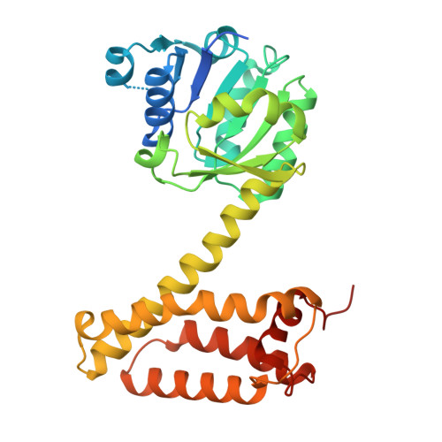 | |
UniProt | |||||
Find proteins for I0BDU9 (Paenibacillus mucilaginosus K02) Explore I0BDU9 Go to UniProtKB: I0BDU9 | |||||
Entity Groups | |||||
| Sequence Clusters | 30% Identity50% Identity70% Identity90% Identity95% Identity100% Identity | ||||
| UniProt Group | I0BDU9 | ||||
Sequence AnnotationsExpand | |||||
| |||||
| Length ( Å ) | Angle ( ˚ ) |
|---|---|
| a = 133.771 | α = 90 |
| b = 133.771 | β = 90 |
| c = 62.23 | γ = 120 |
| Software Name | Purpose |
|---|---|
| PHENIX | refinement |
| HKL-3000 | data reduction |
| HKL-3000 | data scaling |
| MOLREP | phasing |
| Funding Organization | Location | Grant Number |
|---|---|---|
| Shanghai University of Medicine & Health Sciences | China | -- |
RCSB PDB (citation) is hosted by
RCSB PDB is a member of the

