


Structure of DNA ligase A from Mycobacterium tuberculosis bound to NAD
Delker, S.L., Abendroth, J., Lorimer, D.D., Horanyi, P.S., Edwards, T.E.To be published.
Experimental Data Snapshot
Entity ID: 1 | |||||
|---|---|---|---|---|---|
| Molecule | Chains | Sequence Length | Organism | Details | Image |
| DNA ligase A | 336 | Mycobacterium tuberculosis H37Rv | Mutation(s): 0 Gene Names: ligA, lig, Rv3014c, MTV012.28c EC: 6.5.1.2 | 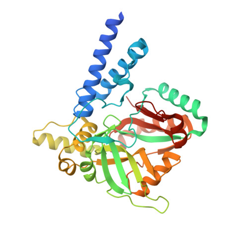 | |
UniProt | |||||
Find proteins for P9WNV1 (Mycobacterium tuberculosis (strain ATCC 25618 / H37Rv)) Explore P9WNV1 Go to UniProtKB: P9WNV1 | |||||
Entity Groups | |||||
| Sequence Clusters | 30% Identity50% Identity70% Identity90% Identity95% Identity100% Identity | ||||
| UniProt Group | P9WNV1 | ||||
Sequence AnnotationsExpand | |||||
| |||||
| Ligands 4 Unique | |||||
|---|---|---|---|---|---|
| ID | Chains | Name / Formula / InChI Key | 2D Diagram | 3D Interactions | |
| NAD Query on NAD | E [auth A], J [auth B], P [auth C], T [auth D] | NICOTINAMIDE-ADENINE-DINUCLEOTIDE C21 H27 N7 O14 P2 BAWFJGJZGIEFAR-NNYOXOHSSA-N |  | ||
| MPD Query on MPD | I [auth A], N [auth B], O [auth B], S [auth C], X [auth D] | (4S)-2-METHYL-2,4-PENTANEDIOL C6 H14 O2 SVTBMSDMJJWYQN-YFKPBYRVSA-N |  | ||
| CA Query on CA | H [auth A], M [auth B] | CALCIUM ION Ca BHPQYMZQTOCNFJ-UHFFFAOYSA-N |  | ||
| MG Query on MG | F [auth A] G [auth A] K [auth B] L [auth B] Q [auth C] | MAGNESIUM ION Mg JLVVSXFLKOJNIY-UHFFFAOYSA-N |  | ||
| Length ( Å ) | Angle ( ˚ ) |
|---|---|
| a = 50.94 | α = 96.22 |
| b = 63.81 | β = 89.55 |
| c = 120.73 | γ = 111.97 |
| Software Name | Purpose |
|---|---|
| PHENIX | refinement |
| XSCALE | data scaling |
| PDB_EXTRACT | data extraction |
| XDS | data reduction |
| PHASER | phasing |
RCSB PDB (citation) is hosted by
RCSB PDB is a member of the

