7F5J

a mutant of an enzyme from Viola yedoensis


Experimental Data Snapshot

  • Method: X-RAY DIFFRACTION
  • Resolution: 1.59 Å
  • R-Value Free: 0.193 
  • R-Value Work: 0.163 

wwPDB Validation   3D Report Full Report


Ligand Structure Quality Assessment 


This is version 2.1 of the entry. See complete history


Literature

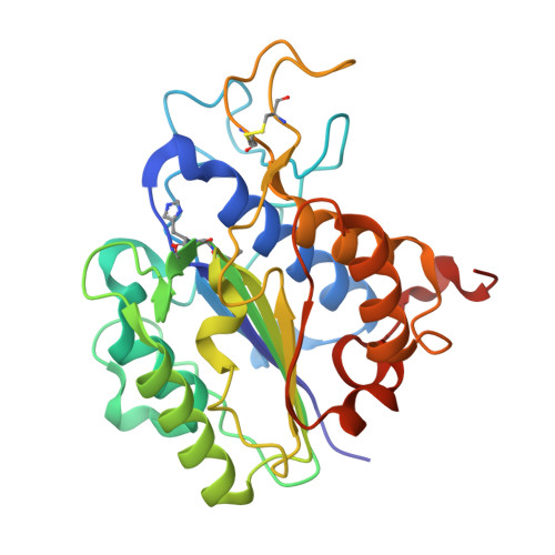
Structural basis for proenzyme maturation, substrate recognition, and ligation by a hyperactive peptide asparaginyl ligase.

Hu, S.El Sahili, A.Kishore, S.Wong, Y.H.Hemu, X.Goh, B.C.Zhipei, S.Wang, Z.Tam, J.P.Liu, C.F.Lescar, J.

(2022) Plant Cell 34: 4936-4949

  • DOI: https://doi.org/10.1093/plcell/koac281
  • Primary Citation of Related Structures:  
    7F5J, 7F5P, 7F5Q, 7FA0

  • PubMed Abstract: 

    Peptide ligases are versatile enzymes that can be utilized for precise protein conjugation for bioengineering applications. Hyperactive peptide asparaginyl ligases (PALs), such as butelase-1, belong to a small class of enzymes from cyclotide-producing plants that can perform site-specific, rapid ligation reactions after a target peptide asparagine/aspartic acid (Asx) residue binds to the active site of the ligase. How PALs specifically recognize their polypeptide substrates has remained elusive, especially at the prime binding side of the enzyme. Here we report crystal structures that capture VyPAL2, a catalytically efficient PAL from Viola yedoensis, in an activated state, with and without a bound substrate. The bound structure shows one ligase with the N-terminal polypeptide tail from another ligase molecule trapped at its active site, revealing how Asx inserts in the enzyme's S1 pocket and why a hydrophobic residue is required at the P2' position. Besides illustrating the anchoring role played by P1 and P2' residues, these results uncover a role for the Gatekeeper residue at the surface of the S2 pocket in shifting the nonprime portion of the substrate and, as a result, the activity toward ligation or hydrolysis. These results suggest a picture for proenzyme maturation in the vacuole and will inform the rational design of peptide ligases with tailored specificities.


  • Organizational Affiliation

    School of Biological Sciences, Nanyang Technological University, Singapore City, 637551, Singapore.


Macromolecules
Find similar proteins by:  (by identity cutoff)  |  3D Structure
Entity ID: 1
MoleculeChains Sequence LengthOrganismDetailsImage
Peptide Asparaginyl Ligases
A, B
282Viola philippicaMutation(s): 1 
UniProt
Find proteins for A0A509GV09 (Viola philippica)
Explore A0A509GV09 
Go to UniProtKB:  A0A509GV09
Entity Groups  
Sequence Clusters30% Identity50% Identity70% Identity90% Identity95% Identity100% Identity
UniProt GroupA0A509GV09
Sequence Annotations
Expand
  • Reference Sequence
Oligosaccharides

Help

Entity ID: 2
MoleculeChains Length2D Diagram Glycosylation3D Interactions
alpha-L-fucopyranose-(1-6)-2-acetamido-2-deoxy-beta-D-glucopyranose
C
2N-Glycosylation
Glycosylation Resources
GlyTouCan:  G86851RC
GlyCosmos:  G86851RC
GlyGen:  G86851RC
Entity ID: 3
MoleculeChains Length2D Diagram Glycosylation3D Interactions
2-acetamido-2-deoxy-beta-D-glucopyranose-(1-4)-2-acetamido-2-deoxy-beta-D-glucopyranose
D
2N-Glycosylation
Glycosylation Resources
GlyTouCan:  G42666HT
GlyCosmos:  G42666HT
GlyGen:  G42666HT
Small Molecules
Ligands 3 Unique
IDChains Name / Formula / InChI Key2D Diagram3D Interactions
NAG
Query on NAG

Download Ideal Coordinates CCD File 
E [auth A],
H [auth A],
N [auth B],
P [auth B]
2-acetamido-2-deoxy-beta-D-glucopyranose
C8 H15 N O6
OVRNDRQMDRJTHS-FMDGEEDCSA-N
GOL
Query on GOL

Download Ideal Coordinates CCD File 
J [auth A],
L [auth A],
M [auth A],
O [auth B]
GLYCEROL
C3 H8 O3
PEDCQBHIVMGVHV-UHFFFAOYSA-N
EDO
Query on EDO

Download Ideal Coordinates CCD File 
F [auth A]
G [auth A]
I [auth A]
K [auth A]
Q [auth B]
F [auth A],
G [auth A],
I [auth A],
K [auth A],
Q [auth B],
R [auth B]
1,2-ETHANEDIOL
C2 H6 O2
LYCAIKOWRPUZTN-UHFFFAOYSA-N
Experimental Data & Validation

Experimental Data

  • Method: X-RAY DIFFRACTION
  • Resolution: 1.59 Å
  • R-Value Free: 0.193 
  • R-Value Work: 0.163 
  • Space Group: P 21 21 21
Unit Cell:
Length ( Å )Angle ( ˚ )
a = 76.27α = 90
b = 78.98β = 90
c = 92.12γ = 90
Software Package:
Software NamePurpose
BUSTERrefinement
autoPROCdata reduction
XDSdata scaling
MOLREPphasing

Structure Validation

View Full Validation Report


Ligand Structure Quality Assessment 


Entry History & Funding Information

Deposition Data


Funding OrganizationLocationGrant Number
Ministry of Education (MoE, Singapore)SingaporeMOE2016-T3-1-003

Revision History  (Full details and data files)

  • Version 1.0: 2022-06-29
    Type: Initial release
  • Version 1.1: 2022-11-30
    Changes: Database references
  • Version 1.2: 2022-12-14
    Changes: Database references
  • Version 2.0: 2023-11-15
    Changes: Atomic model, Data collection
  • Version 2.1: 2023-11-29
    Changes: Refinement description