7B75

Cryo-EM Structure of Human Thyroglobulin


Experimental Data Snapshot

  • Method: ELECTRON MICROSCOPY
  • Resolution: 3.20 Å
  • Aggregation State: PARTICLE 
  • Reconstruction Method: SINGLE PARTICLE 

wwPDB Validation   3D Report Full Report


This is version 1.1 of the entry. See complete history


Literature

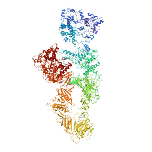
Cryo-EM structure of native human thyroglobulin.

Adaixo, R.Steiner, E.M.Righetto, R.D.Schmidt, A.Stahlberg, H.Taylor, N.M.I.

(2022) Nat Commun 13: 61-61

  • DOI: https://doi.org/10.1038/s41467-021-27693-8
  • Primary Citation of Related Structures:  
    7B75

  • PubMed Abstract: 

    The thyroglobulin (TG) protein is essential to thyroid hormone synthesis, plays a vital role in the regulation of metabolism, development and growth and serves as intraglandular iodine storage. Its architecture is conserved among vertebrates. Synthesis of triiodothyronine (T 3 ) and thyroxine (T 4 ) hormones depends on the conformation, iodination and post-translational modification of TG. Although structural information is available on recombinant and deglycosylated endogenous human thyroglobulin (hTG) from patients with goiters, the structure of native, fully glycosylated hTG remained unknown. Here, we present the cryo-electron microscopy structure of native and fully glycosylated hTG from healthy thyroid glands to 3.2 Å resolution. The structure provides detailed information on hormonogenic and glycosylation sites. We employ liquid chromatography-mass spectrometry (LC-MS) to validate these findings as well as other post-translational modifications and proteolytic cleavage sites. Our results offer insights into thyroid hormonogenesis of native hTG and provide a fundamental understanding of clinically relevant mutations.


  • Organizational Affiliation

    Center for Cellular Imaging and NanoAnalytics, Biozentrum, University of Basel, Mattenstrasse 26, 4058, Basel, Switzerland.


Macromolecules
Find similar proteins by:  (by identity cutoff)  |  3D Structure
Entity ID: 1
MoleculeChains Sequence LengthOrganismDetailsImage
Thyroglobulin
A, B
2,768Homo sapiensMutation(s): 0 
UniProt & NIH Common Fund Data Resources
Find proteins for P01266 (Homo sapiens)
Explore P01266 
Go to UniProtKB:  P01266
PHAROS:  P01266
GTEx:  ENSG00000042832 
Entity Groups  
Sequence Clusters30% Identity50% Identity70% Identity90% Identity95% Identity100% Identity
UniProt GroupP01266
Sequence Annotations
Expand
  • Reference Sequence
Oligosaccharides

Help

Entity ID: 2
MoleculeChains Length2D Diagram Glycosylation3D Interactions
2-acetamido-2-deoxy-beta-D-glucopyranose-(1-4)-2-acetamido-2-deoxy-beta-D-glucopyranose
C, D, F, I, J
C, D, F, I, J, L
2N-Glycosylation
Glycosylation Resources
GlyTouCan:  G42666HT
GlyCosmos:  G42666HT
GlyGen:  G42666HT
Entity ID: 3
MoleculeChains Length2D Diagram Glycosylation3D Interactions
beta-D-mannopyranose-(1-4)-2-acetamido-2-deoxy-beta-D-glucopyranose-(1-4)-2-acetamido-2-deoxy-beta-D-glucopyranose
E, K
3N-Glycosylation
Glycosylation Resources
GlyTouCan:  G15407YE
GlyCosmos:  G15407YE
GlyGen:  G15407YE
Entity ID: 4
MoleculeChains Length2D Diagram Glycosylation3D Interactions
alpha-D-mannopyranose-(1-3)-beta-D-mannopyranose-(1-4)-2-acetamido-2-deoxy-beta-D-glucopyranose-(1-4)-2-acetamido-2-deoxy-beta-D-glucopyranose
G, M
4N-Glycosylation
Glycosylation Resources
GlyTouCan:  G81315DD
GlyCosmos:  G81315DD
GlyGen:  G81315DD
Entity ID: 5
MoleculeChains Length2D Diagram Glycosylation3D Interactions
alpha-D-mannopyranose-(1-3)-alpha-D-mannopyranose-(1-3)-[alpha-D-mannopyranose-(1-6)]beta-D-mannopyranose-(1-4)-2-acetamido-2-deoxy-beta-D-glucopyranose-(1-4)-2-acetamido-2-deoxy-beta-D-glucopyranose
H, N
6N-Glycosylation
Glycosylation Resources
GlyTouCan:  G42984ZU
GlyCosmos:  G42984ZU
GlyGen:  G42984ZU
Small Molecules
Ligands 1 Unique
IDChains Name / Formula / InChI Key2D Diagram3D Interactions
NAG (Subject of Investigation/LOI)
Query on NAG

Download Ideal Coordinates CCD File 
AA [auth B]
BA [auth B]
CA [auth B]
DA [auth B]
O [auth A]
AA [auth B],
BA [auth B],
CA [auth B],
DA [auth B],
O [auth A],
P [auth A],
Q [auth A],
R [auth A],
S [auth A],
T [auth A],
U [auth A],
V [auth A],
W [auth B],
X [auth B],
Y [auth B],
Z [auth B]
2-acetamido-2-deoxy-beta-D-glucopyranose
C8 H15 N O6
OVRNDRQMDRJTHS-FMDGEEDCSA-N
Experimental Data & Validation

Experimental Data

  • Method: ELECTRON MICROSCOPY
  • Resolution: 3.20 Å
  • Aggregation State: PARTICLE 
  • Reconstruction Method: SINGLE PARTICLE 
EM Software:
TaskSoftware PackageVersion
MODEL REFINEMENTCoot0.9.3
RECONSTRUCTIONRELION3

Structure Validation

View Full Validation Report


Entry History & Funding Information

Deposition Data


Funding OrganizationLocationGrant Number
Swiss National Science FoundationDenmark--
Novo Nordisk FoundationDenmarkNNF14CC0001

Revision History  (Full details and data files)

  • Version 1.0: 2021-12-29
    Type: Initial release
  • Version 1.1: 2022-01-26
    Changes: Database references