6Z3G

Repulsive Guidance Molecule A (RGMA) in complex with Growth Differentiation Factor 5 (GDF5)


Experimental Data Snapshot

  • Method: X-RAY DIFFRACTION
  • Resolution: 2.78 Å
  • R-Value Free: 0.288 
  • R-Value Work: 0.234 
  • R-Value Observed: 0.236 

wwPDB Validation   3D Report Full Report


Ligand Structure Quality Assessment 


This is version 1.3 of the entry. See complete history


Literature

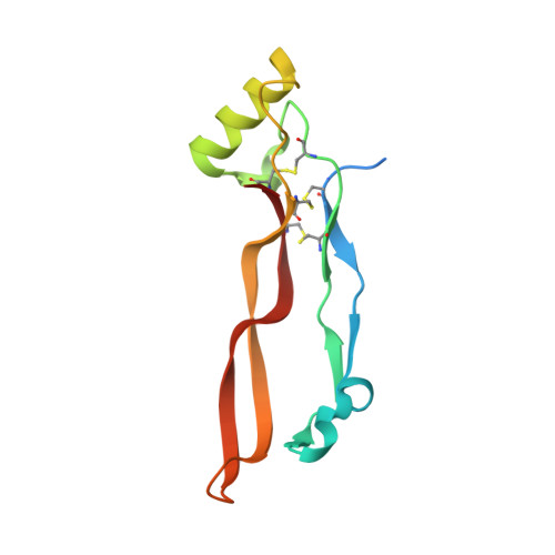
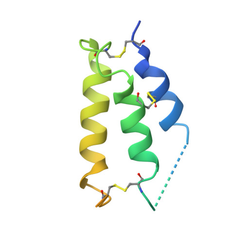
Repulsive guidance molecules lock growth differentiation factor 5 in an inhibitory complex.

Malinauskas, T.Peer, T.V.Bishop, B.Mueller, T.D.Siebold, C.

(2020) Proc Natl Acad Sci U S A 117: 15620-15631

  • DOI: https://doi.org/10.1073/pnas.2000561117
  • Primary Citation of Related Structures:  
    6Z3G, 6Z3H, 6Z3J, 6Z3L, 6Z3M

  • PubMed Abstract: 

    Repulsive guidance molecules (RGMs) are cell surface proteins that regulate the development and homeostasis of many tissues and organs, including the nervous, skeletal, and immune systems. They control fundamental biological processes, such as migration and differentiation by direct interaction with the Neogenin (NEO1) receptor and function as coreceptors for the bone morphogenetic protein (BMP)/growth differentiation factor (GDF) family. We determined crystal structures of all three human RGM family members in complex with GDF5, as well as the ternary NEO1-RGMB-GDF5 assembly. Surprisingly, we show that all three RGMs inhibit GDF5 signaling, which is in stark contrast to RGM-mediated enhancement of signaling observed for other BMPs, like BMP2. Despite their opposite effect on GDF5 signaling, RGMs occupy the BMP type 1 receptor binding site similar to the observed interactions in RGM-BMP2 complexes. In the NEO1-RGMB-GDF5 complex, RGMB physically bridges NEO1 and GDF5, suggesting cross-talk between the GDF5 and NEO1 signaling pathways. Our crystal structures, combined with structure-guided mutagenesis of RGMs and BMP ligands, binding studies, and cellular assays suggest that RGMs inhibit GDF5 signaling by competing with GDF5 type 1 receptors. While our crystal structure analysis and in vitro binding data initially pointed towards a simple competition mechanism between RGMs and type 1 receptors as a possible basis for RGM-mediated GDF5 inhibition, further experiments utilizing BMP2-mimicking GDF5 variants clearly indicate a more complex mechanism that explains how RGMs can act as a functionality-changing switch for two structurally and biochemically similar signaling molecules.


  • Organizational Affiliation

    Division of Structural Biology, Wellcome Centre for Human Genetics, University of Oxford, OX3 7BN Oxford, United Kingdom; tomas@strubi.ox.ac.uk mueller@biozentrum.uni-wuerzburg.de christian@strubi.ox.ac.uk.


Macromolecules
Find similar proteins by:  (by identity cutoff)  |  3D Structure
Entity ID: 1
MoleculeChains Sequence LengthOrganismDetailsImage
Growth/differentiation factor 5117Homo sapiensMutation(s): 0 
Gene Names: GDF5BMP14CDMP1
UniProt & NIH Common Fund Data Resources
Find proteins for P43026 (Homo sapiens)
Explore P43026 
Go to UniProtKB:  P43026
PHAROS:  P43026
GTEx:  ENSG00000125965 
Entity Groups  
Sequence Clusters30% Identity50% Identity70% Identity90% Identity95% Identity100% Identity
UniProt GroupP43026
Sequence Annotations
Expand
  • Reference Sequence
Find similar proteins by:  (by identity cutoff)  |  3D Structure
Entity ID: 2
MoleculeChains Sequence LengthOrganismDetailsImage
Repulsive guidance molecule A106Homo sapiensMutation(s): 0 
Gene Names: RGMARGM
UniProt & NIH Common Fund Data Resources
Find proteins for Q96B86 (Homo sapiens)
Explore Q96B86 
Go to UniProtKB:  Q96B86
PHAROS:  Q96B86
GTEx:  ENSG00000182175 
Entity Groups  
Sequence Clusters30% Identity50% Identity70% Identity90% Identity95% Identity100% Identity
UniProt GroupQ96B86
Sequence Annotations
Expand
  • Reference Sequence
Small Molecules
Ligands 1 Unique
IDChains Name / Formula / InChI Key2D Diagram3D Interactions
CIT (Subject of Investigation/LOI)
Query on CIT

Download Ideal Coordinates CCD File 
C [auth A]CITRIC ACID
C6 H8 O7
KRKNYBCHXYNGOX-UHFFFAOYSA-N
Experimental Data & Validation

Experimental Data

  • Method: X-RAY DIFFRACTION
  • Resolution: 2.78 Å
  • R-Value Free: 0.288 
  • R-Value Work: 0.234 
  • R-Value Observed: 0.236 
  • Space Group: P 62 2 2
Unit Cell:
Length ( Å )Angle ( ˚ )
a = 97.65α = 90
b = 97.65β = 90
c = 99.82γ = 120
Software Package:
Software NamePurpose
PHENIXrefinement
XDSdata reduction
Aimlessdata scaling
PHASERphasing

Structure Validation

View Full Validation Report


Ligand Structure Quality Assessment 


Entry History & Funding Information

Deposition Data


Funding OrganizationLocationGrant Number
Cancer Research UKUnited KingdomC20724/A14414
Cancer Research UKUnited KingdomC20724/A26752
European Research Council (ERC)United Kingdom647278
German Research Foundation (DFG)GermanyMU1095/3-2
German Research Foundation (DFG)GermanyMU1095/5-1
Wellcome TrustUnited Kingdom203852/Z/16/2
Human Frontier Science Program (HFSP)United KingdomLT000021/2014-L

Revision History  (Full details and data files)

  • Version 1.0: 2020-07-01
    Type: Initial release
  • Version 1.1: 2020-07-08
    Changes: Database references
  • Version 1.2: 2020-07-15
    Changes: Database references
  • Version 1.3: 2024-01-24
    Changes: Data collection, Database references, Derived calculations, Refinement description