6TP5

Crystal structure of human Transmembrane prolyl 4-hydroxylase


Experimental Data Snapshot

  • Method: X-RAY DIFFRACTION
  • Resolution: 2.25 Å
  • R-Value Free: 0.221 
  • R-Value Work: 0.181 
  • R-Value Observed: 0.183 

wwPDB Validation   3D Report Full Report


Ligand Structure Quality Assessment 


This is version 1.3 of the entry. See complete history


Literature

Structure of transmembrane prolyl 4-hydroxylase reveals unique organization of EF and dioxygenase domains.

Myllykoski, M.Sutinen, A.Koski, M.K.Kallio, J.P.Raasakka, A.Myllyharju, J.Wierenga, R.K.Koivunen, P.

(2020) J Biol Chem 296: 100197-100197

  • DOI: https://doi.org/10.1074/jbc.RA120.016542
  • Primary Citation of Related Structures:  
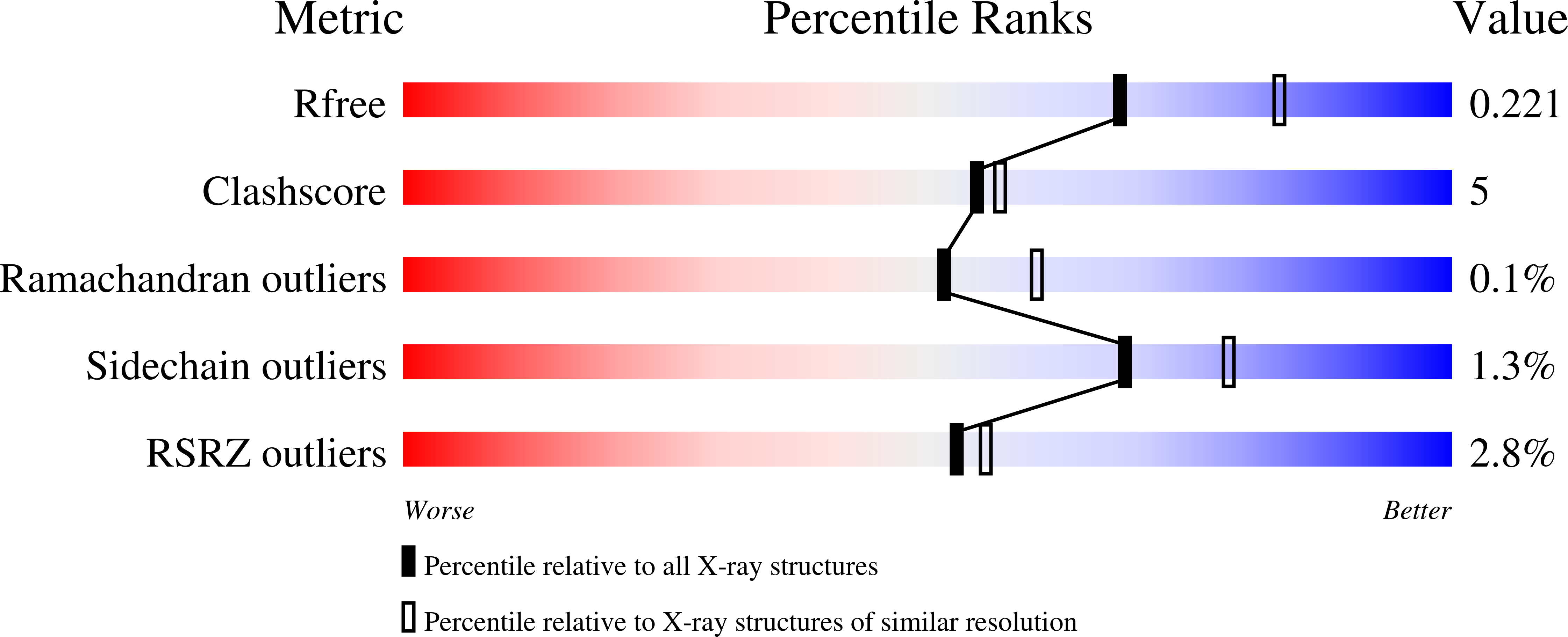
    6TP5

  • PubMed Abstract: 

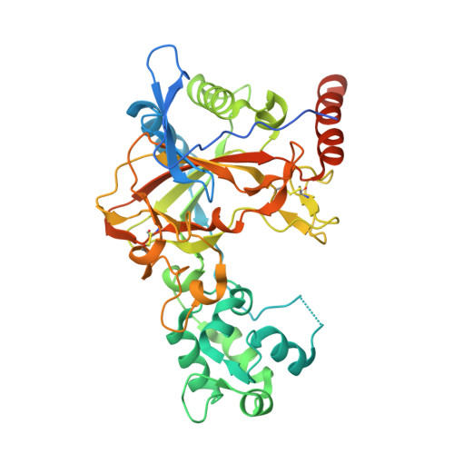
    Prolyl 4-hydroxylases (P4Hs) catalyze post-translational hydroxylation of peptidyl proline residues. In addition to collagen P4Hs and hypoxia-inducible factor P4Hs, a third P4H-the poorly characterized endoplasmic reticulum-localized transmembrane prolyl 4-hydroxylase (P4H-TM)-is found in animals. P4H-TM variants are associated with the familiar neurological HIDEA syndrome, but how these variants might contribute to disease is unknown. Here, we explored this question in a structural and functional analysis of soluble human P4H-TM. The crystal structure revealed an EF domain with two Ca 2+ -binding motifs inserted within the catalytic domain. A substrate-binding groove was formed between the EF domain and the conserved core of the catalytic domain. The proximity of the EF domain to the active site suggests that Ca 2+ binding is relevant to the catalytic activity. Functional analysis demonstrated that Ca 2+ -binding affinity of P4H-TM is within the range of physiological Ca 2+ concentration in the endoplasmic reticulum. P4H-TM was found both as a monomer and a dimer in the solution, but the monomer-dimer equilibrium was not regulated by Ca 2+ . The catalytic site contained bound Fe 2+ and N-oxalylglycine, which is an analogue of the cosubstrate 2-oxoglutarate. Comparison with homologous P4H structures complexed with peptide substrates showed that the substrate-interacting residues and the lid structure that folds over the substrate are conserved in P4H-TM, whereas the extensive loop structures that surround the substrate-binding groove, generating a negative surface potential, are different. Analysis of the structure suggests that the HIDEA variants cause loss of P4H-TM function. In conclusion, P4H-TM shares key structural elements with other P4Hs while having a unique EF domain.


  • Organizational Affiliation

    Biocenter Oulu, Faculty of Biochemistry and Molecular Medicine, University of Oulu, Oulu, Finland; Oulu Center for Cell-Matrix Research, University of Oulu, Oulu, Finland.


Macromolecules
Find similar proteins by:  (by identity cutoff)  |  3D Structure
Entity ID: 1
MoleculeChains Sequence LengthOrganismDetailsImage
Transmembrane prolyl 4-hydroxylase
A, B
421Homo sapiensMutation(s): 0 
Gene Names: P4HTMPH4
EC: 1.14.11.29
UniProt & NIH Common Fund Data Resources
Find proteins for Q9NXG6 (Homo sapiens)
Explore Q9NXG6 
Go to UniProtKB:  Q9NXG6
PHAROS:  Q9NXG6
GTEx:  ENSG00000178467 
Entity Groups  
Sequence Clusters30% Identity50% Identity70% Identity90% Identity95% Identity100% Identity
UniProt GroupQ9NXG6
Sequence Annotations
Expand
  • Reference Sequence
Oligosaccharides

Help

Entity ID: 2
MoleculeChains Length2D Diagram Glycosylation3D Interactions
2-acetamido-2-deoxy-beta-D-glucopyranose-(1-4)-2-acetamido-2-deoxy-beta-D-glucopyranose
C, E
2N-Glycosylation
Glycosylation Resources
GlyTouCan:  G42666HT
GlyCosmos:  G42666HT
GlyGen:  G42666HT
Entity ID: 3
MoleculeChains Length2D Diagram Glycosylation3D Interactions
alpha-D-mannopyranose-(1-2)-alpha-D-mannopyranose-(1-2)-alpha-D-mannopyranose-(1-3)-[alpha-D-mannopyranose-(1-2)-alpha-D-mannopyranose-(1-6)-[alpha-D-mannopyranose-(1-3)]alpha-D-mannopyranose-(1-6)]beta-D-mannopyranose-(1-4)-2-acetamido-2-deoxy-beta-D-glucopyranose-(1-4)-2-acetamido-2-deoxy-beta-D-glucopyranose
D
10N-Glycosylation
Glycosylation Resources
GlyTouCan:  G40702WU
GlyCosmos:  G40702WU
GlyGen:  G40702WU
Entity ID: 4
MoleculeChains Length2D Diagram Glycosylation3D Interactions
beta-D-mannopyranose-(1-2)-alpha-D-mannopyranose-(1-2)-alpha-D-mannopyranose-(1-3)-[alpha-D-mannopyranose-(1-2)-alpha-D-mannopyranose-(1-6)-[alpha-D-mannopyranose-(1-3)]alpha-D-mannopyranose-(1-6)]beta-D-mannopyranose-(1-4)-2-acetamido-2-deoxy-beta-D-glucopyranose-(1-4)-2-acetamido-2-deoxy-beta-D-glucopyranose
F
10N-Glycosylation
Glycosylation Resources
GlyTouCan:  G46700WH
GlyCosmos:  G46700WH
GlyGen:  G46700WH
Small Molecules
Ligands 6 Unique
IDChains Name / Formula / InChI Key2D Diagram3D Interactions
OGA (Subject of Investigation/LOI)
Query on OGA

Download Ideal Coordinates CCD File 
G [auth A],
S [auth B]
N-OXALYLGLYCINE
C4 H5 N O5
BIMZLRFONYSTPT-UHFFFAOYSA-N
GLY
Query on GLY

Download Ideal Coordinates CCD File 
M [auth A],
N [auth A]
GLYCINE
C2 H5 N O2
DHMQDGOQFOQNFH-UHFFFAOYSA-N
TBU
Query on TBU

Download Ideal Coordinates CCD File 
I [auth A]
J [auth A]
K [auth A]
L [auth A]
U [auth B]
I [auth A],
J [auth A],
K [auth A],
L [auth A],
U [auth B],
V [auth B],
W [auth B],
X [auth B]
TERTIARY-BUTYL ALCOHOL
C4 H10 O
DKGAVHZHDRPRBM-UHFFFAOYSA-N
FE2 (Subject of Investigation/LOI)
Query on FE2

Download Ideal Coordinates CCD File 
H [auth A],
T [auth B]
FE (II) ION
Fe
CWYNVVGOOAEACU-UHFFFAOYSA-N
CA (Subject of Investigation/LOI)
Query on CA

Download Ideal Coordinates CCD File 
O [auth A],
P [auth A],
Y [auth B],
Z [auth B]
CALCIUM ION
Ca
BHPQYMZQTOCNFJ-UHFFFAOYSA-N
CL
Query on CL

Download Ideal Coordinates CCD File 
Q [auth A],
R [auth A]
CHLORIDE ION
Cl
VEXZGXHMUGYJMC-UHFFFAOYSA-M
Experimental Data & Validation

Experimental Data

  • Method: X-RAY DIFFRACTION
  • Resolution: 2.25 Å
  • R-Value Free: 0.221 
  • R-Value Work: 0.181 
  • R-Value Observed: 0.183 
  • Space Group: P 31
Unit Cell:
Length ( Å )Angle ( ˚ )
a = 92.079α = 90
b = 92.079β = 90
c = 129.5γ = 120
Software Package:
Software NamePurpose
PHENIXrefinement
XDSdata reduction
XDSdata scaling
PHASERphasing
Cootmodel building

Structure Validation

View Full Validation Report


Ligand Structure Quality Assessment 


Entry History & Funding Information

Deposition Data


Funding OrganizationLocationGrant Number
Academy of FinlandFinland308009
Jane and Aatos Erkko FoundationFinland--
Sigrid Juselius FoundationFinland--

Revision History  (Full details and data files)

  • Version 1.0: 2020-12-23
    Type: Initial release
  • Version 1.1: 2020-12-30
    Changes: Database references
  • Version 1.2: 2021-07-21
    Changes: Database references
  • Version 1.3: 2024-01-24
    Changes: Data collection, Database references, Refinement description