6Q5U

High resolution electron cryo-microscopy structure of the bacteriophage PR772


Experimental Data Snapshot

  • Method: ELECTRON MICROSCOPY
  • Resolution: 2.75 Å
  • Aggregation State: PARTICLE 
  • Reconstruction Method: SINGLE PARTICLE 

wwPDB Validation   3D Report Full Report


This is version 1.4 of the entry. See complete history


Literature

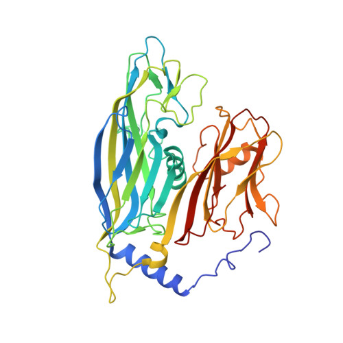
Electron cryo-microscopy of Bacteriophage PR772 reveals the elusive vertex complex and the capsid architecture.

Reddy, H.K.Hajdu, J.Carroni, M.Svenda, M.

(2019) Elife 8

  • DOI: https://doi.org/10.7554/eLife.48496
  • Primary Citation of Related Structures:  
    6Q5U

  • PubMed Abstract: 

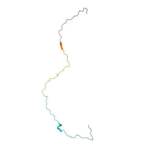
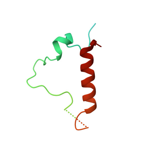
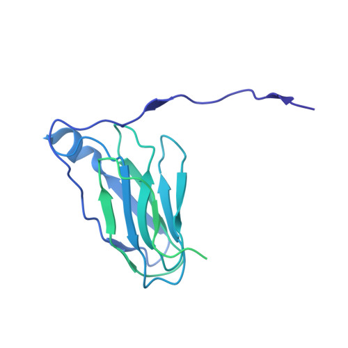
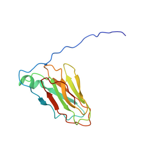
    Bacteriophage PR772, a member of the Tectiviridae family, has a 70 nm diameter icosahedral protein capsid that encapsulates a lipid membrane, dsDNA, and various internal proteins. An icosahedrally averaged CryoEM reconstruction of the wild-type virion and a localized reconstruction of the vertex region reveal the composition and the structure of the vertex complex along with new protein conformations that play a vital role in maintaining the capsid architecture of the virion. The overall resolution of the virion is 2.75 Å, while the resolution of the protein capsid is 2.3 Å. The conventional penta-symmetron formed by the capsomeres is replaced by a large vertex complex in the pseudo T = 25 capsid. All the vertices contain the host-recognition protein, P5; two of these vertices show the presence of the receptor-binding protein, P2. The 3D structure of the vertex complex shows interactions with the viral membrane, indicating a possible mechanism for viral infection.


  • Organizational Affiliation

    Department of Cell and Molecular Biology, Laboratory of Molecular Biophysics, Uppsala University, Uppsala, Sweden.


Macromolecules
Find similar proteins by:  (by identity cutoff)  |  3D Structure
Entity ID: 1
MoleculeChains Sequence LengthOrganismDetailsImage
Major Capsid Protein (P3)
A, B, C, D, E
A, B, C, D, E, F, G, H, I, J, K, L
395Enterobacteria phage PR772Mutation(s): 0 
UniProt
Find proteins for Q6EDX0 (Enterobacteria phage PR772)
Explore Q6EDX0 
Go to UniProtKB:  Q6EDX0
Entity Groups  
Sequence Clusters30% Identity50% Identity70% Identity90% Identity95% Identity100% Identity
UniProt GroupQ6EDX0
Sequence Annotations
Expand
  • Reference Sequence
Find similar proteins by:  (by identity cutoff)  |  3D Structure
Entity ID: 2
MoleculeChains Sequence LengthOrganismDetailsImage
Minor Capsid Protein (P30)84Enterobacteria phage PR772Mutation(s): 0 
UniProt
Find proteins for Q6EDW3 (Enterobacteria phage PR772)
Explore Q6EDW3 
Go to UniProtKB:  Q6EDW3
Entity Groups  
Sequence Clusters30% Identity50% Identity70% Identity90% Identity95% Identity100% Identity
UniProt GroupQ6EDW3
Sequence Annotations
Expand
  • Reference Sequence
Find similar proteins by:  (by identity cutoff)  |  3D Structure
Entity ID: 3
MoleculeChains Sequence LengthOrganismDetailsImage
Infectivity Protein (P16)117Enterobacteria phage PR772Mutation(s): 0 
UniProt
Find proteins for Q6EDW1 (Enterobacteria phage PR772)
Explore Q6EDW1 
Go to UniProtKB:  Q6EDW1
Entity Groups  
Sequence Clusters30% Identity50% Identity70% Identity90% Identity95% Identity100% Identity
UniProt GroupQ6EDW1
Sequence Annotations
Expand
  • Reference Sequence
Find similar proteins by:  (by identity cutoff)  |  3D Structure
Entity ID: 4
MoleculeChains Sequence LengthOrganismDetailsImage
Spike protein
O, P, Q
340Enterobacteria phage PR772Mutation(s): 0 
UniProt
Find proteins for Q6EDX8 (Enterobacteria phage PR772)
Explore Q6EDX8 
Go to UniProtKB:  Q6EDX8
Entity Groups  
Sequence Clusters30% Identity50% Identity70% Identity90% Identity95% Identity100% Identity
UniProt GroupQ6EDX8
Sequence Annotations
Expand
  • Reference Sequence
Find similar proteins by:  (by identity cutoff)  |  3D Structure
Entity ID: 5
MoleculeChains Sequence LengthOrganismDetailsImage
Penton protein
R, S
126Enterobacteria phage PR772Mutation(s): 0 
UniProt
Find proteins for Q6EDY0 (Enterobacteria phage PR772)
Explore Q6EDY0 
Go to UniProtKB:  Q6EDY0
Entity Groups  
Sequence Clusters30% Identity50% Identity70% Identity90% Identity95% Identity100% Identity
UniProt GroupQ6EDY0
Sequence Annotations
Expand
  • Reference Sequence
Experimental Data & Validation

Experimental Data

  • Method: ELECTRON MICROSCOPY
  • Resolution: 2.75 Å
  • Aggregation State: PARTICLE 
  • Reconstruction Method: SINGLE PARTICLE 
EM Software:
TaskSoftware PackageVersion
MODEL REFINEMENTPHENIXdev-3360

Structure Validation

View Full Validation Report


Entry History & Funding Information

Deposition Data


Funding OrganizationLocationGrant Number
Swedish Research CouncilSweden628-20081109822-2010-6157, 822-2012-5260 and 828-2012-108
Knut and Alice Wallenberg FoundationSwedenKAW-2011.081
European Research CouncilSwedenERC-291602

Revision History  (Full details and data files)

  • Version 1.0: 2019-06-19
    Type: Initial release
  • Version 1.1: 2019-07-03
    Changes: Data collection
  • Version 1.2: 2019-09-25
    Changes: Data collection, Database references
  • Version 1.3: 2019-11-06
    Changes: Data collection, Refinement description
  • Version 1.4: 2019-12-18
    Changes: Other