6ON2

Lon Protease from Yersinia pestis with Y2853 substrate

  • Classification: HYDROLASE
  • Organism(s): Yersinia pestis
  • Expression System: Escherichia coli
  • Mutation(s): No 

  • Deposited: 2019-04-19 Released: 2019-05-01 
  • Deposition Author(s): Shin, M., Asmita, A., Puchades, C., Adjei, E., Wiseman, R.L., Karzai, A.W., Lander, G.C.
  • Funding Organization(s): National Institutes of Health/National Institute of Biomedical Imaging and Bioengineering (NIH/NIBIB), National Institutes of Health/National Institute of Neurological Disorders and Stroke (NIH/NINDS), National Institutes of Health/National Institute Of Allergy and Infectious Diseases (NIH/NIAID), National Institutes of Health/National Institute on Aging (NIH/NIA), National Institutes of Health/Office of the Director

Experimental Data Snapshot

  • Method: ELECTRON MICROSCOPY
  • Resolution: 3.00 Å
  • Aggregation State: PARTICLE 
  • Reconstruction Method: SINGLE PARTICLE 

wwPDB Validation   3D Report Full Report


This is version 1.3 of the entry. See complete history


Literature

Structural basis for distinct operational modes and protease activation in AAA+ protease Lon.

Shin, M.Puchades, C.Asmita, A.Puri, N.Adjei, E.Wiseman, R.L.Karzai, A.W.Lander, G.C.

(2020) Sci Adv 6: eaba8404-eaba8404

  • DOI: https://doi.org/10.1126/sciadv.aba8404
  • Primary Citation of Related Structures:  
    6ON2, 6V11

  • PubMed Abstract: 

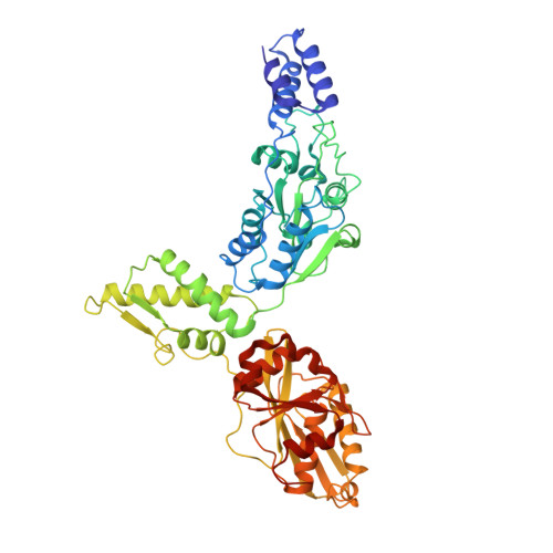
    Substrate-bound structures of AAA+ protein translocases reveal a conserved asymmetric spiral staircase architecture wherein a sequential ATP hydrolysis cycle drives hand-over-hand substrate translocation. However, this configuration is unlikely to represent the full conformational landscape of these enzymes, as biochemical studies suggest distinct conformational states depending on the presence or absence of substrate. Here, we used cryo-electron microscopy to determine structures of the Yersinia pestis Lon AAA+ protease in the absence and presence of substrate, uncovering the mechanistic basis for two distinct operational modes. In the absence of substrate, Lon adopts a left-handed, "open" spiral organization with autoinhibited proteolytic active sites. Upon the addition of substrate, Lon undergoes a reorganization to assemble an enzymatically active, right-handed "closed" conformer with active protease sites. These findings define the mechanistic principles underlying the operational plasticity required for processing diverse protein substrates.


  • Organizational Affiliation

    Department of Integrative Structural and Computational Biology, The Scripps Research Institute, La Jolla, CA 92037, USA.


Macromolecules
Find similar proteins by:  (by identity cutoff)  |  3D Structure
Entity ID: 1
MoleculeChains Sequence LengthOrganismDetailsImage
ATP-dependent protease La
A, B, C, D, E
A, B, C, D, E, F
524Yersinia pestisMutation(s): 0 
Gene Names: lonYP_0776EGT45_05820NCTC144_01047
EC: 3.4.21.53
UniProt
Find proteins for A0A5P8YJ65 (Yersinia pestis)
Explore A0A5P8YJ65 
Go to UniProtKB:  A0A5P8YJ65
Entity Groups  
Sequence Clusters30% Identity50% Identity70% Identity90% Identity95% Identity100% Identity
UniProt GroupA0A5P8YJ65
Sequence Annotations
Expand
  • Reference Sequence

Find similar proteins by:  Sequence   |   3D Structure  

Entity ID: 2
MoleculeChains Sequence LengthOrganismDetailsImage
Bound Y2853 Substrate7Yersinia pestisMutation(s): 0 
Sequence Annotations
Expand
  • Reference Sequence
Small Molecules
Ligands 3 Unique
IDChains Name / Formula / InChI Key2D Diagram3D Interactions
ATP
Query on ATP

Download Ideal Coordinates CCD File 
H [auth A],
J [auth B],
L [auth C],
N [auth D]
ADENOSINE-5'-TRIPHOSPHATE
C10 H16 N5 O13 P3
ZKHQWZAMYRWXGA-KQYNXXCUSA-N
ADP
Query on ADP

Download Ideal Coordinates CCD File 
P [auth E],
R [auth F]
ADENOSINE-5'-DIPHOSPHATE
C10 H15 N5 O10 P2
XTWYTFMLZFPYCI-KQYNXXCUSA-N
MG
Query on MG

Download Ideal Coordinates CCD File 
I [auth A],
K [auth B],
M [auth C],
O [auth D],
Q [auth E]
MAGNESIUM ION
Mg
JLVVSXFLKOJNIY-UHFFFAOYSA-N
Experimental Data & Validation

Experimental Data

  • Method: ELECTRON MICROSCOPY
  • Resolution: 3.00 Å
  • Aggregation State: PARTICLE 
  • Reconstruction Method: SINGLE PARTICLE 
EM Software:
TaskSoftware PackageVersion
RECONSTRUCTIONRELION2.0b
MODEL REFINEMENTPHENIX1.11.1

Structure Validation

View Full Validation Report


Entry History & Funding Information

Deposition Data


Funding OrganizationLocationGrant Number
National Institutes of Health/National Institute of Biomedical Imaging and Bioengineering (NIH/NIBIB)United StatesDP2EB020402
National Institutes of Health/National Institute of Neurological Disorders and Stroke (NIH/NINDS)United StatesNS095892
National Institutes of Health/National Institute Of Allergy and Infectious Diseases (NIH/NIAID)United StatesAI-127533
National Institutes of Health/National Institute on Aging (NIH/NIA)United StatesAG061697
National Institutes of Health/Office of the DirectorUnited StatesS10OD021634

Revision History  (Full details and data files)

  • Version 1.0: 2019-05-01
    Type: Initial release
  • Version 1.1: 2019-12-11
    Changes: Author supporting evidence
  • Version 1.2: 2020-12-02
    Changes: Database references, Derived calculations
  • Version 1.3: 2024-03-20
    Changes: Data collection, Database references