6LIV

Crystal structure of Tyrosine decarboxylase in complex with PLP

  • Classification: LYASE
  • Organism(s): Papaver somniferum
  • Expression System: Escherichia coli
  • Mutation(s): No 

  • Deposited: 2019-12-13 Released: 2020-02-12 
  • Deposition Author(s): Wang, H., Yu, J., Yao, M.
  • Funding Organization(s): Ministry of Education, Culture, Sports, Science and Technology (Japan), Japan Society for the Promotion of Science (JSPS)

Experimental Data Snapshot

  • Method: X-RAY DIFFRACTION
  • Resolution: 2.31 Å
  • R-Value Free: 0.250 
  • R-Value Work: 0.227 
  • R-Value Observed: 0.228 

wwPDB Validation   3D Report Full Report


This is version 1.0 of the entry. See complete history


Literature

Crystal structures clarify cofactor binding of plant tyrosine decarboxylase.

Wang, H.Yu, J.Satoh, Y.Nakagawa, Y.Tanaka, R.Kato, K.Yao, M.

(2019) Biochem Biophys Res Commun 

  • DOI: https://doi.org/10.1016/j.bbrc.2019.12.077
  • Primary Citation of Related Structures:  
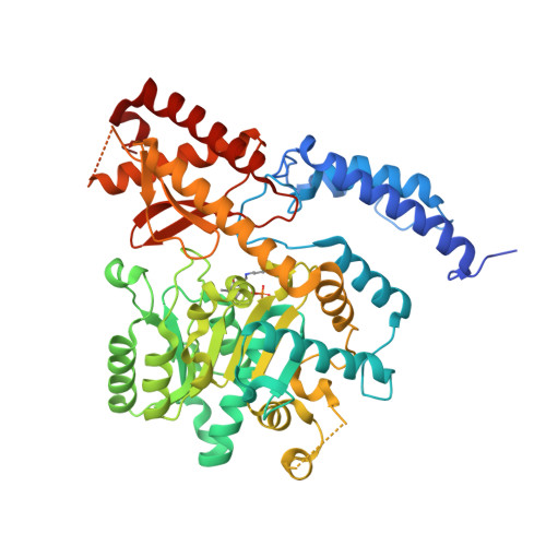
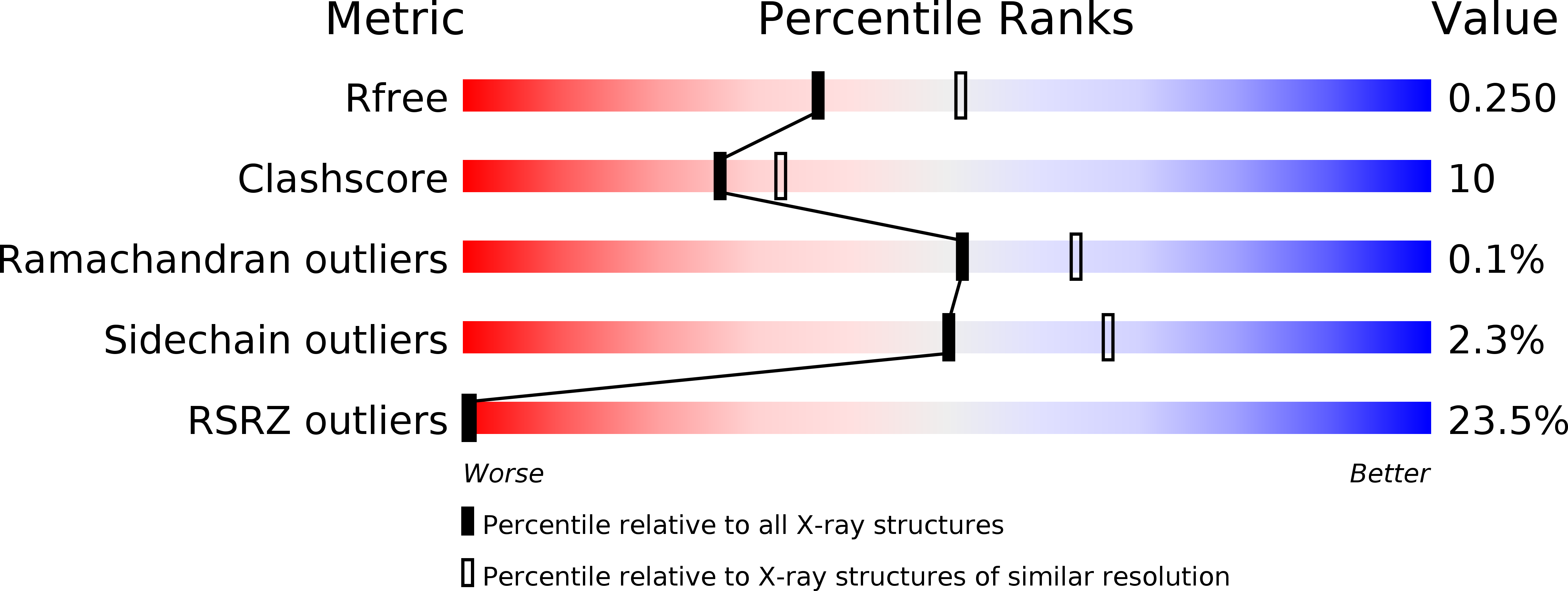
    6LIU, 6LIV

  • PubMed Abstract: 

    Plant tyrosine decarboxylase (TyrDC) is a group II pyridoxal 5'-phosphate (PLP)-dependent decarboxylase that mainly catalyzes the decarboxylation of tyrosine to tyramine. This is biologically important for diverting essential primary metabolites into secondary metabolic pathways. Intensive studies have characterized the effective of PLP-binding and the substrate specificity of mammalian 3,4-dihydroxyphenyl-l-alanine (Dopa) decarboxylases, a member of group II PLP-dependent decarboxylase. However, the characteristics of PLP binding and substrate specificity of plant TyrDCs remain unknown. In this study, we focus on the PLP binding manner, and determined the crystal structures of the apo and PLP binding form of type II TyrDC from Papaver somniferum (PsTyrDCII and PsTyrDCII-PLP). The structures showed that, unlike mammalian Dopa decarboxylase, the binding of PLP does not induce distinct conformational changes of PsTyrDCII regarding the overall structure, but the PLP binding pocket displays conformational changes at Phe124, His203 and Thr262. Combining structural comparation and the obtained biochemical findings, it is demonstrated that PsTyrDCII does not binds PLP tightly. Such characteristics of PLP binding may be required by its catalytic reaction and substrate binding. The activity of TyrDC probably regulated by the concentration of PLP in cells.


  • Organizational Affiliation

    Graduate School of Life Science, Hokkaido University, Sapporo, Hokkaido, 060-0810, Japan.


Macromolecules
Find similar proteins by:  (by identity cutoff)  |  3D Structure
Entity ID: 1
MoleculeChains Sequence LengthOrganismDetailsImage
Tyrosine/DOPA decarboxylase 2
A, B, C, D, E
A, B, C, D, E, F
531Papaver somniferumMutation(s): 0 
Gene Names: TYDC2
EC: 4.1.1.28 (PDB Primary Data), 4.1.1.25 (PDB Primary Data)
UniProt
Find proteins for P54769 (Papaver somniferum)
Explore P54769 
Go to UniProtKB:  P54769
Entity Groups  
Sequence Clusters30% Identity50% Identity70% Identity90% Identity95% Identity100% Identity
UniProt GroupP54769
Sequence Annotations
Expand
  • Reference Sequence
Experimental Data & Validation

Experimental Data

  • Method: X-RAY DIFFRACTION
  • Resolution: 2.31 Å
  • R-Value Free: 0.250 
  • R-Value Work: 0.227 
  • R-Value Observed: 0.228 
  • Space Group: P 21 2 21
Unit Cell:
Length ( Å )Angle ( ˚ )
a = 117.911α = 90
b = 180.414β = 90
c = 218.057γ = 90
Software Package:
Software NamePurpose
XDSdata reduction
XDSdata scaling
PHASERphasing
PHENIXmodel building
PHENIXrefinement

Structure Validation

View Full Validation Report


Entry History & Funding Information

Deposition Data


Funding OrganizationLocationGrant Number
Ministry of Education, Culture, Sports, Science and Technology (Japan)Japan17H05424
Japan Society for the Promotion of Science (JSPS)Japan19H04633

Revision History  (Full details and data files)

  • Version 1.0: 2020-02-12
    Type: Initial release