6KKJ

Crystal structure of Drug:Proton Antiporter-1 (DHA1) Family SotB, in the inward open conformation


Experimental Data Snapshot

  • Method: X-RAY DIFFRACTION
  • Resolution: 3.38 Å
  • R-Value Free: 0.287 
  • R-Value Work: 0.249 
  • R-Value Observed: 0.253 

wwPDB Validation   3D Report Full Report


Ligand Structure Quality Assessment 


This is version 1.2 of the entry. See complete history


Literature

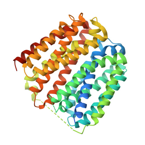
Visualizing the nonlinear changes of a drug-proton antiporter from inward-open to occluded state.

Xiao, Q.Sun, B.Zhou, Y.Wang, C.Guo, L.He, J.Deng, D.

(2021) Biochem Biophys Res Commun 534: 272-278

  • DOI: https://doi.org/10.1016/j.bbrc.2020.11.096
  • Primary Citation of Related Structures:  
    6KKI, 6KKJ, 6KKK, 6KKL

  • PubMed Abstract: 

    Drug-proton antiporters (DHA) play an important role in multi-drug resistance, utilizing the proton-motive force to drive the expulsion of toxic molecules, including antibiotics and drugs. DHA transporters belong to the major facilitator superfamily (MFS), members of which deliver substrates by utilizing the alternating access model of transport. However, the transport process is still elusive. Here, we report the structures of SotB, a member of DHA1 family (TCDB: 2.A.1.2) from Escherichia coli. Four crystal structures of SotB were captured in different conformations, including substrate-bound occluded, inward-facing, and inward-open states. Comparisons between the four structures reveal nonlinear rigid-body movements of alternating access during the state transition from inward-open to occluded conformation. This work not only reveals the conformational dynamics of SotB but also deepens our understanding of the alternating access mechanism of MFS transporters.


  • Organizational Affiliation

    From the Division of Obstetrics, Key Laboratory of Birth Defects and Related Disease of Women and Children of MOE, State Key Laboratory of Biotherapy, West China Second Hospital, Sichuan University, Chengdu, 610041, China.


Macromolecules
Find similar proteins by:  (by identity cutoff)  |  3D Structure
Entity ID: 1
MoleculeChains Sequence LengthOrganismDetailsImage
Sugar efflux transporterA [auth B]423Escherichia coli K-12Mutation(s): 0 
Gene Names: sotBydeAb1528JW1521
Membrane Entity: Yes 
UniProt
Find proteins for P31122 (Escherichia coli (strain K12))
Explore P31122 
Go to UniProtKB:  P31122
Entity Groups  
Sequence Clusters30% Identity50% Identity70% Identity90% Identity95% Identity100% Identity
UniProt GroupP31122
Sequence Annotations
Expand
  • Reference Sequence
Small Molecules
Ligands 1 Unique
IDChains Name / Formula / InChI Key2D Diagram3D Interactions
BNG
Query on BNG

Download Ideal Coordinates CCD File 
B,
C [auth B]
nonyl beta-D-glucopyranoside
C15 H30 O6
QFAPUKLCALRPLH-UXXRCYHCSA-N
Experimental Data & Validation

Experimental Data

  • Method: X-RAY DIFFRACTION
  • Resolution: 3.38 Å
  • R-Value Free: 0.287 
  • R-Value Work: 0.249 
  • R-Value Observed: 0.253 
  • Space Group: P 21 21 21
Unit Cell:
Length ( Å )Angle ( ˚ )
a = 69.227α = 90
b = 89.15β = 90
c = 90.007γ = 90
Software Package:
Software NamePurpose
PHENIXrefinement
HKL-2000data reduction
HKL-2000data scaling
PHASERphasing
PDB_EXTRACTdata extraction

Structure Validation

View Full Validation Report


Ligand Structure Quality Assessment 


Entry History & Funding Information

Deposition Data


Funding OrganizationLocationGrant Number
National Institutes of Health/National Human Genome Research Institute (NIH/NHGRI)ChinaYFA052700
National Science Foundation (NSF, China)ChinaJQ007

Revision History  (Full details and data files)

  • Version 1.0: 2020-07-29
    Type: Initial release
  • Version 1.1: 2021-03-31
    Changes: Database references, Structure summary
  • Version 1.2: 2023-11-22
    Changes: Data collection, Database references, Refinement description