6JTG

Structural insights into G domain dimerization and pathogenic mutations of OPA1

  • Classification: HYDROLASE
  • Organism(s): Homo sapiens
  • Expression System: Escherichia coli
  • Mutation(s): No 

  • Deposited: 2019-04-11 Released: 2020-04-22 
  • Deposition Author(s): Yan, L., Hu, J.
  • Funding Organization(s): National Natural Science Foundation of China, Ministry of Science and Technology (China)

Experimental Data Snapshot

  • Method: X-RAY DIFFRACTION
  • Resolution: 2.40 Å
  • R-Value Free: 0.244 
  • R-Value Work: 0.199 
  • R-Value Observed: 0.204 

wwPDB Validation   3D Report Full Report


This is version 1.2 of the entry. See complete history


Literature

Structural insights into G domain dimerization and pathogenic mutation of OPA1.

Yu, C.Zhao, J.Yan, L.Qi, Y.Guo, X.Lou, Z.Hu, J.Rao, Z.

(2020) J Cell Biol 219

  • DOI: https://doi.org/10.1083/jcb.201907098
  • Primary Citation of Related Structures:  
    6JTG

  • PubMed Abstract: 

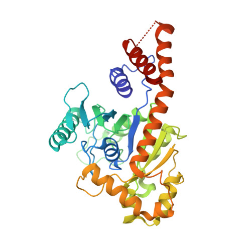
    The fusion of mammalian inner mitochondrial membranes (IMMs) is mediated by dynamin-like GTPase OPA1. Mutations in human OPA1 cause optic atrophy, but the molecular basis for membrane fusion and pathogenesis is not clear. Here, we determined the crystal structure of the minimal GTPase domain (MGD) of human OPA1. A three-helix bundle (HB) domain including two helices extending from the GTPase (G) domain and the last helix of OPA1 tightly associates with the G domain. In the presence of GDP and BeF3-, OPA1-MGD forms a dimer, the interface of which is critical for the maintenance of mitochondrial morphology. The catalytic core of OPA1 possesses unique features that are not present in other dynamin-like proteins. Biochemical experiments revealed that OPA1-MGD forms nucleotide-dependent dimers, which is important for membrane-stimulated GTP hydrolysis, and an N-terminal extension mediates nucleotide-independent dimerization that facilitates efficient membrane association. Our results suggest a multifaceted assembly of OPA1 and explain the effect of most OPA1 mutations on optic atrophy.


  • Organizational Affiliation

    College of Life Sciences and State Key Laboratory of Medicinal Chemical Biology, Nankai University, Tianjin, China.


Macromolecules
Find similar proteins by:  (by identity cutoff)  |  3D Structure
Entity ID: 1
MoleculeChains Sequence LengthOrganismDetailsImage
Dynamin-like 120 kDa protein, mitochondrial,OPA1 protein350Homo sapiensMutation(s): 0 
Gene Names: OPA1
EC: 3.6.5.5
UniProt & NIH Common Fund Data Resources
Find proteins for O60313 (Homo sapiens)
Explore O60313 
Go to UniProtKB:  O60313
PHAROS:  O60313
GTEx:  ENSG00000198836 
Entity Groups  
Sequence Clusters30% Identity50% Identity70% Identity90% Identity95% Identity100% Identity
UniProt GroupO60313
Sequence Annotations
Expand
  • Reference Sequence
Experimental Data & Validation

Experimental Data

  • Method: X-RAY DIFFRACTION
  • Resolution: 2.40 Å
  • R-Value Free: 0.244 
  • R-Value Work: 0.199 
  • R-Value Observed: 0.204 
  • Space Group: P 43 21 2
Unit Cell:
Length ( Å )Angle ( ˚ )
a = 77.89α = 90
b = 77.89β = 90
c = 171.547γ = 90
Software Package:
Software NamePurpose
PHENIXrefinement
PHENIXphasing
PHENIXmodel building

Structure Validation

View Full Validation Report


Entry History & Funding Information

Deposition Data

  • Released Date: 2020-04-22 
  • Deposition Author(s): Yan, L., Hu, J.

Funding OrganizationLocationGrant Number
National Natural Science Foundation of ChinaChina20171301816
Ministry of Science and Technology (China)China2017YFC0840302

Revision History  (Full details and data files)

  • Version 1.0: 2020-04-22
    Type: Initial release
  • Version 1.1: 2020-05-20
    Changes: Database references
  • Version 1.2: 2024-03-27
    Changes: Data collection, Database references, Derived calculations