6H64

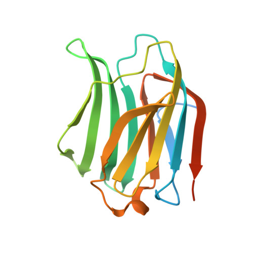
Crystal structure of the CRD-SAT


Experimental Data Snapshot

  • Method: X-RAY DIFFRACTION
  • Resolution: 1.80 Å
  • R-Value Free: 0.238 
  • R-Value Work: 0.201 

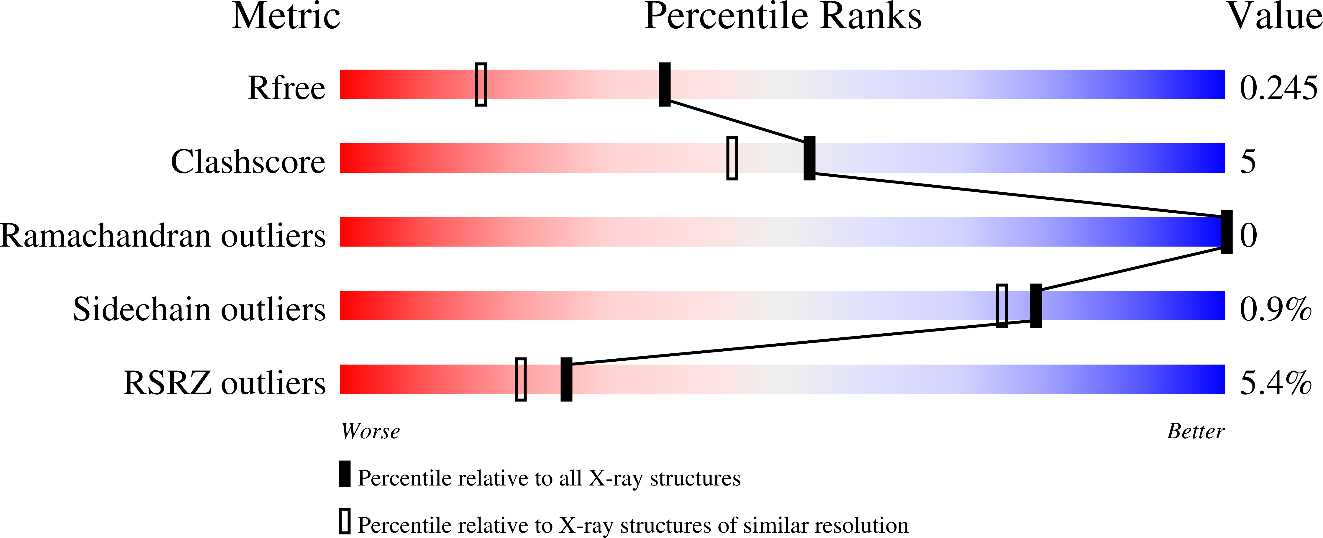
wwPDB Validation   3D Report Full Report


This is version 2.2 of the entry. See complete history


Literature

CRD SAT Generated by pCARGHO: A New Efficient Lectin-Based Affinity Tag Method for Safe, Simple, and Low-Cost Protein Purification.

Kriznik, A.Yelehe-Okouma, M.Lec, J.C.Groshenry, G.Le Cordier, H.Charron, C.Quinternet, M.Mazon, H.Talfournier, F.Boschi-Muller, S.Jouzeau, J.Y.Reboul, P.

(2019) Biotechnol J 14: e1800214-e1800214

  • DOI: https://doi.org/10.1002/biot.201800214
  • Primary Citation of Related Structures:  
    6H64

  • PubMed Abstract: 

    Purification of recombinant proteins remains a bottleneck for downstream processing. The authors engineered a new galectin 3 truncated form (CRD SAT ), functionally and structurally characterized, with preserved solubility and lectinic activity. Taking advantage of these properties, the authors designed an expression vector (pCARGHO), suitable for CRD SAT -tagged protein expression in prokaryotes. CRD SAT binds to lactose-Sepharose with a high specificity and facilitates solubilization of fusion proteins. This tag is structurally stable and can be easily removed from fusion proteins using TEV protease. Furthermore, due to their basic isoelectric point (pI), CRD SAT , and TEV are efficiently eliminated using cationic exchange chromatography. When pI of the protein of interest (POI) and CRD SAT are close, other chromatographic methods are successfully tested. Using CRD SAT tag, the authors purified several proteins from prokaryote and eukaryote origin and demonstrated as examples, the preservation of both Escherichia coli Thioredoxin 1 and human CDC25B cd activities. Overall, yields of proteins obtained after tag removal are about 5-50 mg per litre of bacterial culture. Our purification method displays various advantages described herein that may greatly interest academic laboratories, biotechnology, and pharmaceutical companies.


  • Organizational Affiliation

    Université de Lorraine, CNRS, Ingénierie Moléculaire et Pathologie Articulaire (IMoPA), F-54000, Nancy, France.


Macromolecules
Find similar proteins by:  (by identity cutoff)  |  3D Structure
Entity ID: 1
MoleculeChains Sequence LengthOrganismDetailsImage
Galectin-3
A, B, C, D, E
A, B, C, D, E, F
170Homo sapiensMutation(s): 2 
Gene Names: LGALS3MAC2
UniProt & NIH Common Fund Data Resources
Find proteins for P17931 (Homo sapiens)
Explore P17931 
Go to UniProtKB:  P17931
PHAROS:  P17931
GTEx:  ENSG00000131981 
Entity Groups  
Sequence Clusters30% Identity50% Identity70% Identity90% Identity95% Identity100% Identity
UniProt GroupP17931
Sequence Annotations
Expand
  • Reference Sequence
Oligosaccharides

Help

Entity ID: 2
MoleculeChains Length2D Diagram Glycosylation3D Interactions
beta-D-galactopyranose-(1-4)-alpha-D-glucopyranose
G, H, I, J, K
G, H, I, J, K, L
2N/A
Glycosylation Resources
GlyTouCan:  G88362QR
GlyCosmos:  G88362QR
GlyGen:  G88362QR
Experimental Data & Validation

Experimental Data

  • Method: X-RAY DIFFRACTION
  • Resolution: 1.80 Å
  • R-Value Free: 0.238 
  • R-Value Work: 0.201 
  • Space Group: P 62
Unit Cell:
Length ( Å )Angle ( ˚ )
a = 118.058α = 90
b = 118.058β = 90
c = 137.506γ = 120
Software Package:
Software NamePurpose
REFMACrefinement
XDSdata reduction
XDSdata scaling
PHASERphasing

Structure Validation

View Full Validation Report


Entry History 

Deposition Data

Revision History  (Full details and data files)

  • Version 1.0: 2019-08-14
    Type: Initial release
  • Version 2.0: 2020-07-29
    Type: Remediation
    Reason: Carbohydrate remediation
    Changes: Atomic model, Data collection, Derived calculations, Non-polymer description, Structure summary
  • Version 2.1: 2021-06-16
    Changes: Database references, Structure summary
  • Version 2.2: 2024-01-17
    Changes: Data collection, Database references, Refinement description