6G48

Sporosarcina pasteurii urease inhibited by silver


Experimental Data Snapshot

  • Method: X-RAY DIFFRACTION
  • Resolution: 1.91 Å
  • R-Value Free: 0.209 
  • R-Value Work: 0.167 
  • R-Value Observed: 0.169 

wwPDB Validation   3D Report Full Report


This is version 1.1 of the entry. See complete history


Literature

The structure of urease inactivated by Ag(i): a new paradigm for enzyme inhibition by heavy metals.

Mazzei, L.Cianci, M.Gonzalez Vara, A.Ciurli, S.

(2018) Dalton Trans 47: 8240-8247

  • DOI: https://doi.org/10.1039/c8dt01190g
  • Primary Citation of Related Structures:  
    6G48

  • PubMed Abstract: 

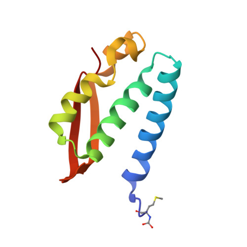
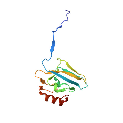
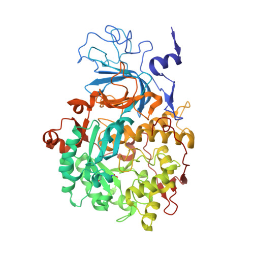
    The nickel-dependent enzyme urease is a virulence factor for a large number of human pathogens, as well as a negative element for the efficiency of soil nitrogen fertilization for crop production. The use of urease inhibitors to contrast these effects requires the knowledge, at the molecular level, of their mode of action. Among these, silver is an efficient antimicrobial agent and an established inhibitor of this enzyme. The 1.91 Å resolution structure of Sporosarcina pasteurii urease inhibited by silver reveals the presence of two Ag(i) ions bound to the largely conserved triad αCys322/αHis323/αMet367: the first two residues are located on the mobile flap that is essential in modulating the size of the active site cavity and the position of key residues for enzyme catalysis, while αMet367 is on a loop facing the flap at the entrance of the active site cavity. The two Ag(i) ions are bridged by the thiolate Sγ atom of αCys322, and are coordinated, respectively, to the Nδ1 atom of the αHis323 imidazole ring and to the Sδ of αMet367. The binding of the Ag(i) ions at the edge of the active site channel supposedly blocks the movement of the flap, inhibiting the catalytic activity of urease. The structure of the silver-inhibited urease allows us to understand and rationalise all previously acquired kinetic and calorimetric data on this phenomenon, but also provides the details of how silver can exert its antimicrobial action with respect to ureolytic bacteria, a step forward against antibiotic-resistant pathogens.


  • Organizational Affiliation

    Laboratory of Bioinorganic Chemistry, Department of Pharmacy and Biotechnology, University of Bologna, Viale G. Fanin 40, I-40127 Bologna, Italy. stefano.ciurli@unibo.it.


Macromolecules
Find similar proteins by:  (by identity cutoff)  |  3D Structure
Entity ID: 1
MoleculeChains Sequence LengthOrganismDetailsImage
Urease subunit gamma100Sporosarcina pasteuriiMutation(s): 0 
EC: 3.5.1.5
UniProt
Find proteins for A0A0H3YGY5 (Sporosarcina pasteurii)
Explore A0A0H3YGY5 
Go to UniProtKB:  A0A0H3YGY5
Entity Groups  
Sequence Clusters30% Identity50% Identity70% Identity90% Identity95% Identity100% Identity
UniProt GroupA0A0H3YGY5
Sequence Annotations
Expand
  • Reference Sequence
Find similar proteins by:  (by identity cutoff)  |  3D Structure
Entity ID: 2
MoleculeChains Sequence LengthOrganismDetailsImage
Urease subunit beta122Sporosarcina pasteuriiMutation(s): 0 
EC: 3.5.1.5
UniProt
Find proteins for P41021 (Sporosarcina pasteurii)
Explore P41021 
Go to UniProtKB:  P41021
Entity Groups  
Sequence Clusters30% Identity50% Identity70% Identity90% Identity95% Identity100% Identity
UniProt GroupP41021
Sequence Annotations
Expand
  • Reference Sequence
Find similar proteins by:  (by identity cutoff)  |  3D Structure
Entity ID: 3
MoleculeChains Sequence LengthOrganismDetailsImage
Urease subunit alpha570Sporosarcina pasteuriiMutation(s): 0 
UniProt
Find proteins for P41020 (Sporosarcina pasteurii)
Explore P41020 
Go to UniProtKB:  P41020
Entity Groups  
Sequence Clusters30% Identity50% Identity70% Identity90% Identity95% Identity100% Identity
UniProt GroupP41020
Sequence Annotations
Expand
  • Reference Sequence
Small Molecules
Ligands 5 Unique
IDChains Name / Formula / InChI Key2D Diagram3D Interactions
AG
Query on AG

Download Ideal Coordinates CCD File 
W [auth C],
X [auth C]
SILVER ION
Ag
FOIXSVOLVBLSDH-UHFFFAOYSA-N
SO4
Query on SO4

Download Ideal Coordinates CCD File 
G [auth A]
I [auth B]
J [auth B]
T [auth C]
U [auth C]
G [auth A],
I [auth B],
J [auth B],
T [auth C],
U [auth C],
V [auth C]
SULFATE ION
O4 S
QAOWNCQODCNURD-UHFFFAOYSA-L
EDO
Query on EDO

Download Ideal Coordinates CCD File 
D [auth A]
E [auth A]
F [auth A]
H [auth B]
N [auth C]
D [auth A],
E [auth A],
F [auth A],
H [auth B],
N [auth C],
O [auth C],
P [auth C],
Q [auth C],
R [auth C],
S [auth C]
1,2-ETHANEDIOL
C2 H6 O2
LYCAIKOWRPUZTN-UHFFFAOYSA-N
NI
Query on NI

Download Ideal Coordinates CCD File 
K [auth C],
L [auth C]
NICKEL (II) ION
Ni
VEQPNABPJHWNSG-UHFFFAOYSA-N
OH
Query on OH

Download Ideal Coordinates CCD File 
M [auth C]HYDROXIDE ION
H O
XLYOFNOQVPJJNP-UHFFFAOYSA-M
Modified Residues  2 Unique
IDChains TypeFormula2D DiagramParent
CXM
Query on CXM
A
L-PEPTIDE LINKINGC6 H11 N O4 SMET
KCX
Query on KCX
C
L-PEPTIDE LINKINGC7 H14 N2 O4LYS
Experimental Data & Validation

Experimental Data

  • Method: X-RAY DIFFRACTION
  • Resolution: 1.91 Å
  • R-Value Free: 0.209 
  • R-Value Work: 0.167 
  • R-Value Observed: 0.169 
  • Space Group: P 63 2 2
Unit Cell:
Length ( Å )Angle ( ˚ )
a = 131.088α = 90
b = 131.088β = 90
c = 189.059γ = 120
Software Package:
Software NamePurpose
REFMACrefinement
XDSdata reduction
Aimlessdata scaling
REFMACphasing

Structure Validation

View Full Validation Report


Entry History 

Deposition Data

Revision History  (Full details and data files)

  • Version 1.0: 2019-01-16
    Type: Initial release
  • Version 1.1: 2024-01-17
    Changes: Data collection, Database references, Derived calculations, Refinement description