6FGA

Crystal structure of TRIM21 E3 ligase, RING domain in complex with its cognate E2 conjugating enzyme UBE2E1


Experimental Data Snapshot

  • Method: X-RAY DIFFRACTION
  • Resolution: 2.82 Å
  • R-Value Free: 0.294 
  • R-Value Work: 0.253 
  • R-Value Observed: 0.255 

wwPDB Validation   3D Report Full Report


This is version 1.1 of the entry. See complete history


Literature

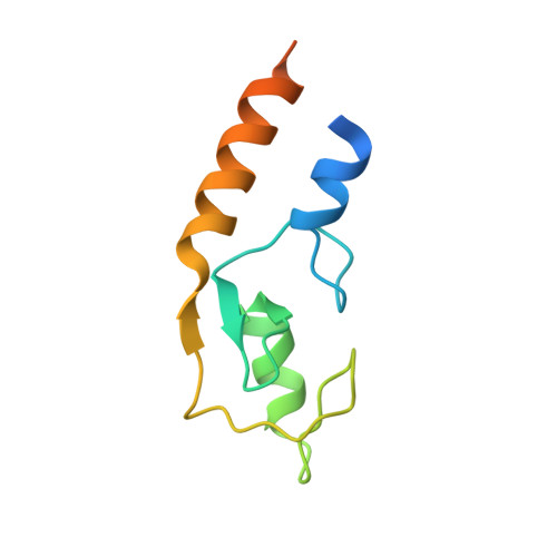
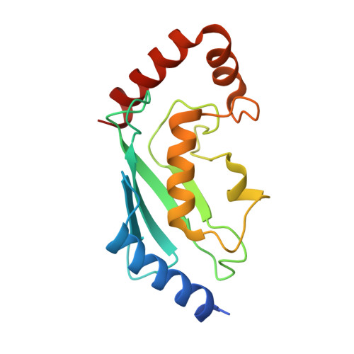
E3 ubiquitin-protein ligase TRIM21-mediated lysine capture by UBE2E1 reveals substrate-targeting mode of a ubiquitin-conjugating E2.

Anandapadamanaban, M.Kyriakidis, N.C.Csizmok, V.Wallenhammar, A.Espinosa, A.C.Ahlner, A.Round, A.R.Trewhella, J.Moche, M.Wahren-Herlenius, M.Sunnerhagen, M.

(2019) J Biol Chem 294: 11404-11419

  • DOI: https://doi.org/10.1074/jbc.RA119.008485
  • Primary Citation of Related Structures:  
    6FGA

  • PubMed Abstract: 

    The E3 ubiquitin-protein ligase TRIM21, of the RING-containing tripartite motif (TRIM) protein family, is a major autoantigen in autoimmune diseases and a modulator of innate immune signaling. Together with ubiquitin-conjugating enzyme E2 E1 (UBE2E1), TRIM21 acts both as an E3 ligase and as a substrate in autoubiquitination. We here report a 2.82-Å crystal structure of the human TRIM21 RING domain in complex with the human E2-conjugating UBE2E1 enzyme, in which a ubiquitin-targeted TRIM21 substrate lysine was captured in the UBE2E1 active site. The structure revealed that the direction of lysine entry is similar to that described for human proliferating cell nuclear antigen (PCNA), a small ubiquitin-like modifier (SUMO)-targeted substrate, and thus differs from the canonical SUMO-targeted substrate entry. In agreement, we found that critical UBE2E1 residues involved in the capture of the TRIM21 substrate lysine are conserved in ubiquitin-conjugating E2s, whereas residues critical for SUMOylation are not conserved. We noted that coordination of the acceptor lysine leads to remodeling of amino acid side-chain interactions between the UBE2E1 active site and the E2-E3 direct interface, including the so-called "linchpin" residue conserved in RING E3s and required for ubiquitination. The findings of our work support the notion that substrate lysine activation of an E2-E3-connecting allosteric path may trigger catalytic activity and contribute to the understanding of specific lysine targeting by ubiquitin-conjugating E2s.


  • Organizational Affiliation

    Department of Physics, Chemistry and Biology, Division of Chemistry, Linköping University, SE-58183 Linköping, Sweden.


Macromolecules
Find similar proteins by:  (by identity cutoff)  |  3D Structure
Entity ID: 1
MoleculeChains Sequence LengthOrganismDetailsImage
E3 ubiquitin-protein ligase TRIM21
A, B, C, D, E
A, B, C, D, E, F, G, H
101Homo sapiensMutation(s): 0 
Gene Names: TRIM21RNF81RO52SSA1
EC: 2.3.2.27
UniProt & NIH Common Fund Data Resources
Find proteins for P19474 (Homo sapiens)
Explore P19474 
Go to UniProtKB:  P19474
PHAROS:  P19474
GTEx:  ENSG00000132109 
Entity Groups  
Sequence Clusters30% Identity50% Identity70% Identity90% Identity95% Identity100% Identity
UniProt GroupP19474
Sequence Annotations
Expand
  • Reference Sequence
Find similar proteins by:  (by identity cutoff)  |  3D Structure
Entity ID: 2
MoleculeChains Sequence LengthOrganismDetailsImage
Ubiquitin-conjugating enzyme E2 E1
I, J, K, L, M
I, J, K, L, M, N, O
158Homo sapiensMutation(s): 0 
Gene Names: UBE2E1UBCH6
EC: 2.3.2.23 (PDB Primary Data), 2.3.2.24 (PDB Primary Data)
UniProt & NIH Common Fund Data Resources
Find proteins for P51965 (Homo sapiens)
Explore P51965 
Go to UniProtKB:  P51965
PHAROS:  P51965
GTEx:  ENSG00000170142 
Entity Groups  
Sequence Clusters30% Identity50% Identity70% Identity90% Identity95% Identity100% Identity
UniProt GroupP51965
Sequence Annotations
Expand
  • Reference Sequence
Small Molecules
Ligands 2 Unique
IDChains Name / Formula / InChI Key2D Diagram3D Interactions
GOL
Query on GOL

Download Ideal Coordinates CCD File 
FA [auth I],
GA [auth I],
HA [auth J],
IA [auth N]
GLYCEROL
C3 H8 O3
PEDCQBHIVMGVHV-UHFFFAOYSA-N
ZN
Query on ZN

Download Ideal Coordinates CCD File 
AA [auth F]
BA [auth G]
CA [auth G]
DA [auth H]
EA [auth H]
AA [auth F],
BA [auth G],
CA [auth G],
DA [auth H],
EA [auth H],
P [auth A],
Q [auth A],
R [auth B],
S [auth B],
T [auth C],
U [auth C],
V [auth D],
W [auth D],
X [auth E],
Y [auth E],
Z [auth F]
ZINC ION
Zn
PTFCDOFLOPIGGS-UHFFFAOYSA-N
Experimental Data & Validation

Experimental Data

  • Method: X-RAY DIFFRACTION
  • Resolution: 2.82 Å
  • R-Value Free: 0.294 
  • R-Value Work: 0.253 
  • R-Value Observed: 0.255 
  • Space Group: C 1 2 1
Unit Cell:
Length ( Å )Angle ( ˚ )
a = 103.835α = 90
b = 95.871β = 93.15
c = 235.097γ = 90
Software Package:
Software NamePurpose
REFMACrefinement
PDB_EXTRACTdata extraction
XDSdata reduction
STARANISOdata scaling
SHELXCDphasing
Cootmodel building

Structure Validation

View Full Validation Report


Entry History & Funding Information

Deposition Data


Funding OrganizationLocationGrant Number
Swedish Research CouncilSweden--

Revision History  (Full details and data files)

  • Version 1.0: 2019-06-12
    Type: Initial release
  • Version 1.1: 2019-08-07
    Changes: Data collection, Database references