6EAC

Pseudomonas syringae SelO

  • Classification: TRANSFERASE
  • Organism(s): Pseudomonas syringae
  • Expression System: Escherichia coli
  • Mutation(s): No 

  • Deposited: 2018-08-02 Released: 2018-10-03 
  • Deposition Author(s): Tomchick, D.R., Tagliabracci, V.S., Sreelatha, A.
  • Funding Organization(s): National Institutes of Health/National Institute of Diabetes and Digestive and Kidney Disease (NIH/NIDDK), National Institutes of Health/National Institute of General Medical Sciences (NIH/NIGMS), Robert A. Welch Foundation, Other government

Experimental Data Snapshot

  • Method: X-RAY DIFFRACTION
  • Resolution: 2.27 Å
  • R-Value Free: 0.212 
  • R-Value Work: 0.182 
  • R-Value Observed: 0.182 

wwPDB Validation   3D Report Full Report


Ligand Structure Quality Assessment 


This is version 1.6 of the entry. See complete history


Literature

Protein AMPylation by an Evolutionarily Conserved Pseudokinase.

Sreelatha, A.Yee, S.S.Lopez, V.A.Park, B.C.Kinch, L.N.Pilch, S.Servage, K.A.Zhang, J.Jiou, J.Karasiewicz-Urbanska, M.Lobocka, M.Grishin, N.V.Orth, K.Kucharczyk, R.Pawlowski, K.Tomchick, D.R.Tagliabracci, V.S.

(2018) Cell 175: 809

  • DOI: https://doi.org/10.1016/j.cell.2018.08.046
  • Primary Citation of Related Structures:  
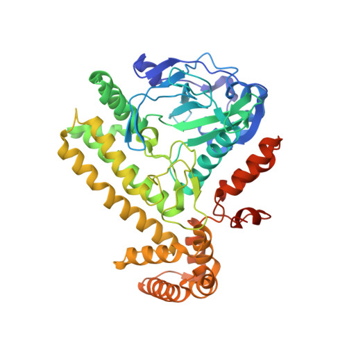
    6EAC

  • PubMed Abstract: 

    Approximately 10% of human protein kinases are believed to be inactive and named pseudokinases because they lack residues required for catalysis. Here, we show that the highly conserved pseudokinase selenoprotein-O (SelO) transfers AMP from ATP to Ser, Thr, and Tyr residues on protein substrates (AMPylation), uncovering a previously unrecognized activity for a member of the protein kinase superfamily. The crystal structure of a SelO homolog reveals a protein kinase-like fold with ATP flipped in the active site, thus providing a structural basis for catalysis. SelO pseudokinases localize to the mitochondria and AMPylate proteins involved in redox homeostasis. Consequently, SelO activity is necessary for the proper cellular response to oxidative stress. Our results suggest that AMPylation may be a more widespread post-translational modification than previously appreciated and that pseudokinases should be analyzed for alternative transferase activities.


  • Organizational Affiliation

    Department of Molecular Biology, University of Texas Southwestern Medical Center, Dallas, TX 75390, USA.


Macromolecules
Find similar proteins by:  (by identity cutoff)  |  3D Structure
Entity ID: 1
MoleculeChains Sequence LengthOrganismDetailsImage
SelO
A, B, C, D
488Pseudomonas syringaeMutation(s): 0 
EC: 2.7.7
UniProt
Find proteins for Q87VB1 (Pseudomonas syringae pv. tomato (strain ATCC BAA-871 / DC3000))
Explore Q87VB1 
Go to UniProtKB:  Q87VB1
Entity Groups  
Sequence Clusters30% Identity50% Identity70% Identity90% Identity95% Identity100% Identity
UniProt GroupQ87VB1
Sequence Annotations
Expand
  • Reference Sequence
Small Molecules
Ligands 7 Unique
IDChains Name / Formula / InChI Key2D Diagram3D Interactions
ANP
Query on ANP

Download Ideal Coordinates CCD File 
CB [auth D],
DA [auth B],
J [auth A],
RA [auth C]
PHOSPHOAMINOPHOSPHONIC ACID-ADENYLATE ESTER
C10 H17 N6 O12 P3
PVKSNHVPLWYQGJ-KQYNXXCUSA-N
PEG
Query on PEG

Download Ideal Coordinates CCD File 
HB [auth D],
KA [auth B],
U [auth A],
UA [auth C],
VA [auth C]
DI(HYDROXYETHYL)ETHER
C4 H10 O3
MTHSVFCYNBDYFN-UHFFFAOYSA-N
EDO
Query on EDO

Download Ideal Coordinates CCD File 
DB [auth D]
EA [auth B]
EB [auth D]
FA [auth B]
FB [auth D]
DB [auth D],
EA [auth B],
EB [auth D],
FA [auth B],
FB [auth D],
GA [auth B],
GB [auth D],
HA [auth B],
IA [auth B],
JA [auth B],
L [auth A],
M [auth A],
N [auth A],
O [auth A],
P [auth A],
Q [auth A],
R [auth A],
S [auth A],
SA [auth C],
T [auth A],
TA [auth C]
1,2-ETHANEDIOL
C2 H6 O2
LYCAIKOWRPUZTN-UHFFFAOYSA-N
ACT
Query on ACT

Download Ideal Coordinates CCD File 
LA [auth B],
V [auth A],
W [auth A],
WA [auth C]
ACETATE ION
C2 H3 O2
QTBSBXVTEAMEQO-UHFFFAOYSA-M
CA
Query on CA

Download Ideal Coordinates CCD File 
AA [auth B]
AB [auth D]
BA [auth B]
F [auth A]
G [auth A]
AA [auth B],
AB [auth D],
BA [auth B],
F [auth A],
G [auth A],
H [auth A],
K [auth A],
NA [auth C],
OA [auth C],
PA [auth C],
Y [auth B],
YA [auth D],
Z [auth B],
ZA [auth D]
CALCIUM ION
Ca
BHPQYMZQTOCNFJ-UHFFFAOYSA-N
CL
Query on CL

Download Ideal Coordinates CCD File 
BB [auth D],
CA [auth B],
I [auth A],
QA [auth C]
CHLORIDE ION
Cl
VEXZGXHMUGYJMC-UHFFFAOYSA-M
MG
Query on MG

Download Ideal Coordinates CCD File 
E [auth A],
MA [auth C],
X [auth B],
XA [auth D]
MAGNESIUM ION
Mg
JLVVSXFLKOJNIY-UHFFFAOYSA-N
Experimental Data & Validation

Experimental Data

  • Method: X-RAY DIFFRACTION
  • Resolution: 2.27 Å
  • R-Value Free: 0.212 
  • R-Value Work: 0.182 
  • R-Value Observed: 0.182 
  • Space Group: I 2 2 2
Unit Cell:
Length ( Å )Angle ( ˚ )
a = 136.174α = 90
b = 158.056β = 90
c = 227.248γ = 90
Software Package:
Software NamePurpose
PHENIXrefinement
HKL-3000data reduction
HKL-3000data scaling
PHENIXmodel building
PDB_EXTRACTdata extraction
PHENIXphasing

Structure Validation

View Full Validation Report


Ligand Structure Quality Assessment 


Entry History & Funding Information

Deposition Data


Funding OrganizationLocationGrant Number
National Institutes of Health/National Institute of Diabetes and Digestive and Kidney Disease (NIH/NIDDK)United StatesR00DK099254
National Institutes of Health/National Institute of General Medical Sciences (NIH/NIGMS)United StatesGM094575
National Institutes of Health/National Institute of General Medical Sciences (NIH/NIGMS)United StatesGM115188
National Institutes of Health/National Institute of Diabetes and Digestive and Kidney Disease (NIH/NIDDK)United StatesT32DK007257-37
National Institutes of Health/National Institute of General Medical Sciences (NIH/NIGMS)United StatesT32GM008203-29
Robert A. Welch FoundationUnited StatesI-1911
Robert A. Welch FoundationUnited StatesI-1505
Robert A. Welch FoundationUnited StatesI-1561
Other governmentUnited StatesRP170674

Revision History  (Full details and data files)

  • Version 1.0: 2018-10-03
    Type: Initial release
  • Version 1.1: 2018-10-10
    Changes: Data collection, Database references, Structure summary
  • Version 1.2: 2018-10-17
    Changes: Data collection, Database references, Structure summary
  • Version 1.3: 2018-10-31
    Changes: Data collection, Database references
  • Version 1.4: 2019-12-25
    Changes: Author supporting evidence
  • Version 1.5: 2020-03-04
    Changes: Data collection
  • Version 1.6: 2024-03-13
    Changes: Data collection, Database references, Derived calculations