6BYK

Structure of 14-3-3 beta/alpha bound to O-ClcNAc peptide


Experimental Data Snapshot

  • Method: X-RAY DIFFRACTION
  • Resolution: 3.00 Å
  • R-Value Free: 0.268 
  • R-Value Work: 0.215 
  • R-Value Observed: 0.220 

wwPDB Validation   3D Report Full Report


Ligand Structure Quality Assessment 


This is version 1.3 of the entry. See complete history


Literature

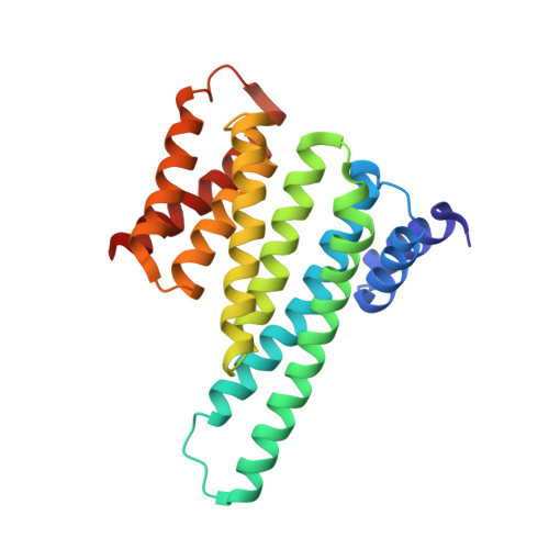
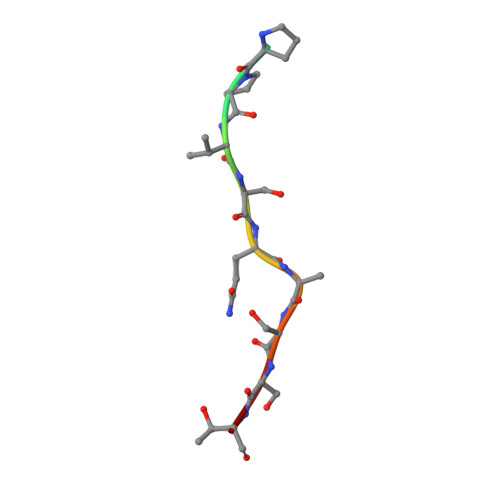
Structural basis of O-GlcNAc recognition by mammalian 14-3-3 proteins.

Toleman, C.A.Schumacher, M.A.Yu, S.H.Zeng, W.Cox, N.J.Smith, T.J.Soderblom, E.J.Wands, A.M.Kohler, J.J.Boyce, M.

(2018) Proc Natl Acad Sci U S A 115: 5956-5961

  • DOI: https://doi.org/10.1073/pnas.1722437115
  • Primary Citation of Related Structures:  
    6BYJ, 6BYK, 6BYL, 6BZD

  • PubMed Abstract: 

    O-GlcNAc is an intracellular posttranslational modification that governs myriad cell biological processes and is dysregulated in human diseases. Despite this broad pathophysiological significance, the biochemical effects of most O-GlcNAcylation events remain uncharacterized. One prevalent hypothesis is that O-GlcNAc moieties may be recognized by "reader" proteins to effect downstream signaling. However, no general O-GlcNAc readers have been identified, leaving a considerable gap in the field. To elucidate O-GlcNAc signaling mechanisms, we devised a biochemical screen for candidate O-GlcNAc reader proteins. We identified several human proteins, including 14-3-3 isoforms, that bind O-GlcNAc directly and selectively. We demonstrate that 14-3-3 proteins bind O-GlcNAc moieties in human cells, and we present the structures of 14-3-3β/α and γ bound to glycopeptides, providing biophysical insights into O-GlcNAc-mediated protein-protein interactions. Because 14-3-3 proteins also bind to phospho-serine and phospho-threonine, they may integrate information from O-GlcNAc and O-phosphate signaling pathways to regulate numerous physiological functions.


  • Organizational Affiliation

    Department of Biochemistry, Duke University School of Medicine, Durham, NC 27710.


Macromolecules
Find similar proteins by:  (by identity cutoff)  |  3D Structure
Entity ID: 1
MoleculeChains Sequence LengthOrganismDetailsImage
14-3-3 protein beta/alpha
A, B, C, D
230Homo sapiensMutation(s): 0 
Gene Names: YWHAB
UniProt & NIH Common Fund Data Resources
Find proteins for P31946 (Homo sapiens)
Explore P31946 
Go to UniProtKB:  P31946
PHAROS:  P31946
GTEx:  ENSG00000166913 
Entity Groups  
Sequence Clusters30% Identity50% Identity70% Identity90% Identity95% Identity100% Identity
UniProt GroupP31946
Sequence Annotations
Expand
  • Reference Sequence

Find similar proteins by:  Sequence   |   3D Structure  

Entity ID: 2
MoleculeChains Sequence LengthOrganismDetailsImage
ATPPVSQASSTT O-GlcNac peptideE [auth G],
F [auth J],
G [auth K],
H [auth R]
12synthetic constructMutation(s): 0 
Entity Groups  
Sequence Clusters30% Identity50% Identity70% Identity90% Identity95% Identity100% Identity
Sequence Annotations
Expand
  • Reference Sequence
Experimental Data & Validation

Experimental Data

  • Method: X-RAY DIFFRACTION
  • Resolution: 3.00 Å
  • R-Value Free: 0.268 
  • R-Value Work: 0.215 
  • R-Value Observed: 0.220 
  • Space Group: P 1 21 1
Unit Cell:
Length ( Å )Angle ( ˚ )
a = 82.67α = 90
b = 71.45β = 111.61
c = 95.759γ = 90
Software Package:
Software NamePurpose
PHENIXrefinement
SCALAdata scaling
MOLREPphasing
PDB_EXTRACTdata extraction
MOSFLMdata reduction

Structure Validation

View Full Validation Report


Ligand Structure Quality Assessment 


Entry History 

Deposition Data

Revision History  (Full details and data files)

  • Version 1.0: 2018-05-09
    Type: Initial release
  • Version 1.1: 2019-05-22
    Changes: Data collection, Database references
  • Version 1.2: 2020-07-29
    Type: Remediation
    Reason: Carbohydrate remediation
    Changes: Data collection, Derived calculations, Structure summary
  • Version 1.3: 2023-10-04
    Changes: Data collection, Database references, Refinement description, Structure summary