6AQ4

CRYSTAL STRUCTURE OF PROTEIN CiTE FROM MYCOBACTERIUM TUBERCULOSIS IN COMPLEX WITH MAGNESIUM, PYRUVATE AND CITRAMALYL-COA


Experimental Data Snapshot

  • Method: X-RAY DIFFRACTION
  • Resolution: 1.82 Å
  • R-Value Free: 0.233 
  • R-Value Work: 0.191 
  • R-Value Observed: 0.192 

wwPDB Validation   3D Report Full Report


Ligand Structure Quality Assessment 


This is version 2.0 of the entry. See complete history


Literature

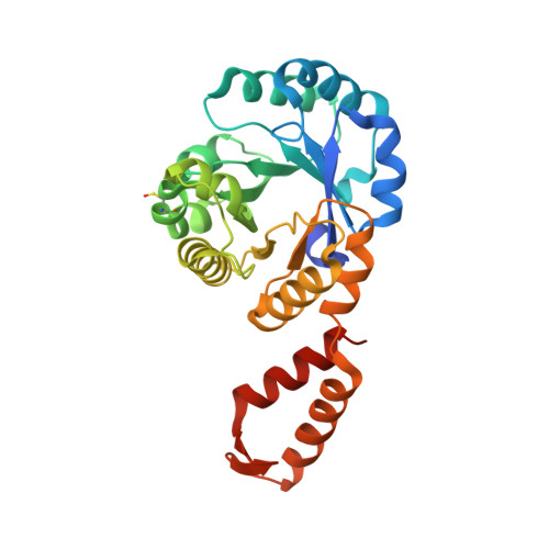
An essential bifunctional enzyme inMycobacterium tuberculosisfor itaconate dissimilation and leucine catabolism.

Wang, H.Fedorov, A.A.Fedorov, E.V.Hunt, D.M.Rodgers, A.Douglas, H.L.Garza-Garcia, A.Bonanno, J.B.Almo, S.C.de Carvalho, L.P.S.

(2019) Proc Natl Acad Sci U S A 116: 15907-15913

  • DOI: https://doi.org/10.1073/pnas.1906606116
  • Primary Citation of Related Structures:  
    6AQ4, 6ARB, 6AS5, 6CHU, 6CJ3, 6CJ4

  • PubMed Abstract: 

    Mycobacterium tuberculosis (Mtb) is the etiological agent of tuberculosis. One-fourth of the global population is estimated to be infected with Mtb, accounting for ∼1.3 million deaths in 2017. As part of the immune response to Mtb infection, macrophages produce metabolites with the purpose of inhibiting or killing the bacterial cell. Itaconate is an abundant host metabolite thought to be both an antimicrobial agent and a modulator of the host inflammatory response. However, the exact mode of action of itaconate remains unclear. Here, we show that Mtb has an itaconate dissimilation pathway and that the last enzyme in this pathway, Rv2498c, also participates in l-leucine catabolism. Our results from phylogenetic analysis, in vitro enzymatic assays, X-ray crystallography, and in vivo Mtb experiments, identified Mtb Rv2498c as a bifunctional β-hydroxyacyl-CoA lyase and that deletion of the rv2498c gene from the Mtb genome resulted in attenuation in a mouse infection model. Altogether, this report describes an itaconate resistance mechanism in Mtb and an l-leucine catabolic pathway that proceeds via an unprecedented ( R )-3-hydroxy-3-methylglutaryl-CoA (HMG-CoA) stereospecific route in nature.


  • Organizational Affiliation

    Mycobacterial Metabolism and Antibiotic Research Laboratory, The Francis Crick Institute, London NW1 1AT, United Kingdom.


Macromolecules
Find similar proteins by:  (by identity cutoff)  |  3D Structure
Entity ID: 1
MoleculeChains Sequence LengthOrganismDetailsImage
Citrate lyase subunit beta-like protein
A, B, C
281Mycobacterium tuberculosis H37RvMutation(s): 0 
Gene Names: citERv2498c
EC: 4.1
UniProt
Find proteins for P9WPE1 (Mycobacterium tuberculosis (strain ATCC 25618 / H37Rv))
Explore P9WPE1 
Go to UniProtKB:  P9WPE1
Entity Groups  
Sequence Clusters30% Identity50% Identity70% Identity90% Identity95% Identity100% Identity
UniProt GroupP9WPE1
Sequence Annotations
Expand
  • Reference Sequence
Small Molecules
Ligands 6 Unique
IDChains Name / Formula / InChI Key2D Diagram3D Interactions
CQM
Query on CQM

Download Ideal Coordinates CCD File 
E [auth A],
L [auth B]
Citramalyl-CoA
C26 H42 N7 O20 P3 S
XYGOWHUIVNMEIA-XBVYHAPZSA-N
PO4
Query on PO4

Download Ideal Coordinates CCD File 
F [auth A],
G [auth A],
M [auth B]
PHOSPHATE ION
O4 P
NBIIXXVUZAFLBC-UHFFFAOYSA-K
GOL
Query on GOL

Download Ideal Coordinates CCD File 
H [auth A],
Q [auth C]
GLYCEROL
C3 H8 O3
PEDCQBHIVMGVHV-UHFFFAOYSA-N
PYR
Query on PYR

Download Ideal Coordinates CCD File 
P [auth C]PYRUVIC ACID
C3 H4 O3
LCTONWCANYUPML-UHFFFAOYSA-N
CL
Query on CL

Download Ideal Coordinates CCD File 
I [auth A],
J [auth A],
N [auth B],
R [auth C],
S [auth C]
CHLORIDE ION
Cl
VEXZGXHMUGYJMC-UHFFFAOYSA-M
MG
Query on MG

Download Ideal Coordinates CCD File 
D [auth A],
K [auth B],
O [auth C]
MAGNESIUM ION
Mg
JLVVSXFLKOJNIY-UHFFFAOYSA-N
Modified Residues  1 Unique
IDChains TypeFormula2D DiagramParent
CSO
Query on CSO
A, B, C
L-PEPTIDE LINKINGC3 H7 N O3 SCYS
Experimental Data & Validation

Experimental Data

  • Method: X-RAY DIFFRACTION
  • Resolution: 1.82 Å
  • R-Value Free: 0.233 
  • R-Value Work: 0.191 
  • R-Value Observed: 0.192 
  • Space Group: C 1 2 1
Unit Cell:
Length ( Å )Angle ( ˚ )
a = 138.541α = 90
b = 88.365β = 108.54
c = 81.252γ = 90
Software Package:
Software NamePurpose
PHENIXrefinement
HKL-2000data reduction
HKL-2000data scaling
PHENIXphasing

Structure Validation

View Full Validation Report


Ligand Structure Quality Assessment 


Entry History 

Deposition Data

Revision History  (Full details and data files)

  • Version 1.0: 2018-08-01
    Type: Initial release
  • Version 1.1: 2019-02-13
    Changes: Data collection, Database references
  • Version 1.2: 2019-07-31
    Changes: Data collection, Database references
  • Version 1.3: 2019-08-21
    Changes: Data collection, Database references
  • Version 1.4: 2023-03-01
    Changes: Database references, Derived calculations, Source and taxonomy, Structure summary
  • Version 1.5: 2023-10-25
    Changes: Data collection, Refinement description
  • Version 2.0: 2023-11-15
    Changes: Atomic model, Data collection, Derived calculations