5ZO7

Kinesin spindle protein Eg5 in complex with STLC-type inhibitor PVEI0138


Experimental Data Snapshot

  • Method: X-RAY DIFFRACTION
  • Resolution: 2.60 Å
  • R-Value Free: 0.283 
  • R-Value Work: 0.225 
  • R-Value Observed: 0.231 

wwPDB Validation   3D Report Full Report


Ligand Structure Quality Assessment 


This is version 1.2 of the entry. See complete history


Literature

Structural and Thermodynamic Basis of the Enhanced Interaction between Kinesin Spindle Protein Eg5 and STLC-type Inhibitors.

Yokoyama, H.Sawada, J.I.Sato, K.Ogo, N.Kamei, N.Ishikawa, Y.Hara, K.Asai, A.Hashimoto, H.

(2018) ACS Omega 3: 12284-12294

  • DOI: https://doi.org/10.1021/acsomega.8b00778
  • Primary Citation of Related Structures:  
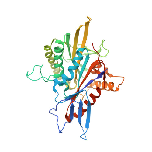
    5ZO7, 5ZO8, 5ZO9

  • PubMed Abstract: 

    For a better understanding of protein-inhibitor interactions, we report structural, thermodynamic, and biological analyses of the interactions between S -trityl-l-cysteine (STLC) derivatives and the motor domain of kinesin spindle protein Eg5. Binding of STLC-type inhibitors to Eg5 was enthalpically driven and entropically unfavorable. The introduction of a para -methoxy substituent in one phenyl ring of STLC enhances its inhibitory activity resulting from a larger enthalpy gain possibly due to the increased shape complementarity. The substituent fits to a recess in the binding pocket. To avoid steric hindrance, the substituted STLC is nudged toward the side opposite to the recess, which enhances the interaction of Eg5 with the remaining part of the inhibitor. Further introduction of an ethylene linkage between two phenyl rings enhances Eg5 inhibitory activity by reducing the loss of entropy in forming the complex. This study provides valuable examples of enhancing protein-inhibitor interactions without forming additional hydrogen bonds.


  • Organizational Affiliation

    Faculty of Pharmaceutical Sciences, Tokyo University of Science, 2641 Yamazaki, Noda, Chiba 278-8510, Japan.


Macromolecules
Find similar proteins by:  (by identity cutoff)  |  3D Structure
Entity ID: 1
MoleculeChains Sequence LengthOrganismDetailsImage
Kinesin-like protein KIF11
A, B
367Homo sapiensMutation(s): 0 
Gene Names: KIF11EG5KNSL1TRIP5
UniProt & NIH Common Fund Data Resources
Find proteins for P52732 (Homo sapiens)
Explore P52732 
Go to UniProtKB:  P52732
PHAROS:  P52732
GTEx:  ENSG00000138160 
Entity Groups  
Sequence Clusters30% Identity50% Identity70% Identity90% Identity95% Identity100% Identity
UniProt GroupP52732
Sequence Annotations
Expand
  • Reference Sequence
Small Molecules
Ligands 4 Unique
IDChains Name / Formula / InChI Key2D Diagram3D Interactions
ADP
Query on ADP

Download Ideal Coordinates CCD File 
C [auth A],
G [auth B]
ADENOSINE-5'-DIPHOSPHATE
C10 H15 N5 O10 P2
XTWYTFMLZFPYCI-KQYNXXCUSA-N
5C5
Query on 5C5

Download Ideal Coordinates CCD File 
E [auth A],
I [auth B]
(2R)-2-azanyl-3-[[2-(4-methoxyphenyl)-2-tricyclo[9.4.0.0^{3,8}]pentadeca-1(11),3,5,7,12,14-hexaenyl]sulfanyl]propanoic acid
C25 H25 N O3 S
FFZDQAVDHIXOIW-QHCPKHFHSA-N
SO4
Query on SO4

Download Ideal Coordinates CCD File 
F [auth A],
J [auth B]
SULFATE ION
O4 S
QAOWNCQODCNURD-UHFFFAOYSA-L
MG
Query on MG

Download Ideal Coordinates CCD File 
D [auth A],
H [auth B]
MAGNESIUM ION
Mg
JLVVSXFLKOJNIY-UHFFFAOYSA-N
Binding Affinity Annotations 
IDSourceBinding Affinity
5C5 BindingDB:  5ZO7 IC50: 14 (nM) from 1 assay(s)
Experimental Data & Validation

Experimental Data

  • Method: X-RAY DIFFRACTION
  • Resolution: 2.60 Å
  • R-Value Free: 0.283 
  • R-Value Work: 0.225 
  • R-Value Observed: 0.231 
  • Space Group: C 1 2 1
Unit Cell:
Length ( Å )Angle ( ˚ )
a = 158.347α = 90
b = 50.58β = 102.2
c = 93.689γ = 90
Software Package:
Software NamePurpose
REFMACrefinement
XDSdata reduction
SCALAdata scaling
MOLREPphasing

Structure Validation

View Full Validation Report


Ligand Structure Quality Assessment 


Entry History & Funding Information

Deposition Data


Funding OrganizationLocationGrant Number
Japan Society for the Promotion of ScienceJapan17K07316

Revision History  (Full details and data files)

  • Version 1.0: 2018-10-10
    Type: Initial release
  • Version 1.1: 2019-09-11
    Changes: Data collection, Database references, Structure summary
  • Version 1.2: 2024-03-27
    Changes: Data collection, Database references, Refinement description