5Z99

Crystal structure of Sialic acid Binding protein from Haemophilus ducreyi with Neu5Ac


Experimental Data Snapshot

  • Method: X-RAY DIFFRACTION
  • Resolution: 1.49 Å
  • R-Value Free: 0.194 
  • R-Value Work: 0.169 
  • R-Value Observed: 0.170 

wwPDB Validation   3D Report Full Report


Ligand Structure Quality Assessment 


This is version 2.1 of the entry. See complete history


Literature

Molecular characterization of the interaction of sialic acid with the periplasmic binding protein fromHaemophilus ducreyi.

Gangi Setty, T.Mowers, J.C.Hobbs, A.G.Maiya, S.P.Syed, S.Munson Jr., R.S.Apicella, M.A.Subramanian, R.

(2018) J Biol Chem 293: 20073-20084

  • DOI: https://doi.org/10.1074/jbc.RA118.005151
  • Primary Citation of Related Structures:  
    5YYB, 5Z99, 5ZA4

  • PubMed Abstract: 

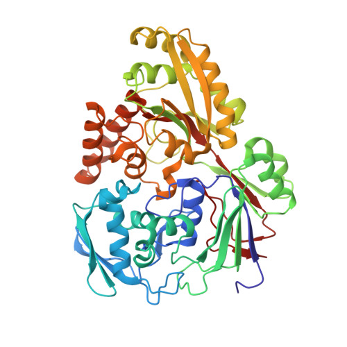
    The primary role of bacterial periplasmic binding proteins is sequestration of essential metabolites present at a low concentration in the periplasm and making them available for active transporters that transfer these ligands into the bacterial cell. The periplasmic binding proteins (SiaPs) from the tripartite ATP-independent periplasmic (TRAP) transport system that transports mammalian host-derived sialic acids have been well studied from different pathogenic bacteria, including Haemophilus influenzae , Fusobacterium nucleatum , Pasteurella multocida , and Vibrio cholerae SiaPs bind the sialic acid N -acetylneuraminic acid (Neu5Ac) with nanomolar affinity by forming electrostatic and hydrogen-bonding interactions. Here, we report the crystal structure of a periplasmic binding protein (SatA) of the ATP-binding cassette (ABC) transport system from the pathogenic bacterium Haemophilus ducreyi The structure of Hd -SatA in the native form and sialic acid-bound forms (with Neu5Ac and N -glycolylneuraminic acid (Neu5Gc)), determined to 2.2, 1.5, and 2.5 Å resolutions, respectively, revealed a ligand-binding site that is very different from those of the SiaPs of the TRAP transport system. A structural comparison along with thermodynamic studies suggested that similar affinities are achieved in the two classes of proteins through distinct mechanisms, one enthalpically driven and the other entropically driven. In summary, our structural and thermodynamic characterization of Hd-SatA reveals that it binds sialic acids with nanomolar affinity and that this binding is an entropically driven process. This information is important for future structure-based drug design against this pathogen and related bacteria.


  • Organizational Affiliation

    From the Institute for Stem Cell Biology and Regenerative Medicine, GKVK Post, Bangalore 560065, India,; the University of Trans-Disciplinary Health Sciences and Technology (TDU), Bengaluru, Karnataka 560064, India.


Macromolecules
Find similar proteins by:  (by identity cutoff)  |  3D Structure
Entity ID: 1
MoleculeChains Sequence LengthOrganismDetailsImage
Putative ABC transporter periplasmic binding protein485[Haemophilus] ducreyi 35000HPMutation(s): 0 
Gene Names: HD_1669
UniProt
Find proteins for Q7VL18 (Haemophilus ducreyi (strain 35000HP / ATCC 700724))
Explore Q7VL18 
Go to UniProtKB:  Q7VL18
Entity Groups  
Sequence Clusters30% Identity50% Identity70% Identity90% Identity95% Identity100% Identity
UniProt GroupQ7VL18
Sequence Annotations
Expand
  • Reference Sequence
Small Molecules
Ligands 1 Unique
IDChains Name / Formula / InChI Key2D Diagram3D Interactions
SLB
Query on SLB

Download Ideal Coordinates CCD File 
B [auth A]N-acetyl-beta-neuraminic acid
C11 H19 N O9
SQVRNKJHWKZAKO-PFQGKNLYSA-N
Binding Affinity Annotations 
IDSourceBinding Affinity
SLB Binding MOAD:  5Z99 Kd: 133 (nM) from 1 assay(s)
Experimental Data & Validation

Experimental Data

  • Method: X-RAY DIFFRACTION
  • Resolution: 1.49 Å
  • R-Value Free: 0.194 
  • R-Value Work: 0.169 
  • R-Value Observed: 0.170 
  • Space Group: P 21 21 21
Unit Cell:
Length ( Å )Angle ( ˚ )
a = 73.368α = 90
b = 89.63β = 90
c = 90.787γ = 90
Software Package:
Software NamePurpose
PHENIXrefinement
PDB_EXTRACTdata extraction
d*TREKdata reduction
Aimlessdata scaling
PHASERphasing

Structure Validation

View Full Validation Report


Ligand Structure Quality Assessment 


Entry History & Funding Information

Deposition Data


Funding OrganizationLocationGrant Number
IndiaSR/SO/BB-19/2010
IndiaBT/IN/Sweden/41/SR/2013

Revision History  (Full details and data files)

  • Version 1.0: 2018-10-24
    Type: Initial release
  • Version 1.1: 2019-01-16
    Changes: Data collection, Database references
  • Version 2.0: 2020-07-29
    Type: Remediation
    Reason: Carbohydrate remediation
    Changes: Atomic model, Data collection, Derived calculations, Structure summary
  • Version 2.1: 2023-11-22
    Changes: Data collection, Database references, Refinement description, Structure summary