5XCV

Crystal structure of NZ-1 Fv-clasp fragment with its antigen peptide


Experimental Data Snapshot

  • Method: X-RAY DIFFRACTION
  • Resolution: 2.14 Å
  • R-Value Free: 0.277 
  • R-Value Work: 0.228 
  • R-Value Observed: 0.231 

wwPDB Validation   3D Report Full Report


This is version 1.2 of the entry. See complete history


Literature

Fv-clasp: An Artificially Designed Small Antibody Fragment with Improved Production Compatibility, Stability, and Crystallizability

Arimori, T.Kitago, Y.Umitsu, M.Fujii, Y.Asaki, R.Tamura-Kawakami, K.Takagi, J.

(2017) Structure 25: 1611-1622.e4

  • DOI: https://doi.org/10.1016/j.str.2017.08.011
  • Primary Citation of Related Structures:  
    5XCQ, 5XCR, 5XCS, 5XCT, 5XCU, 5XCV, 5XCX

  • PubMed Abstract: 

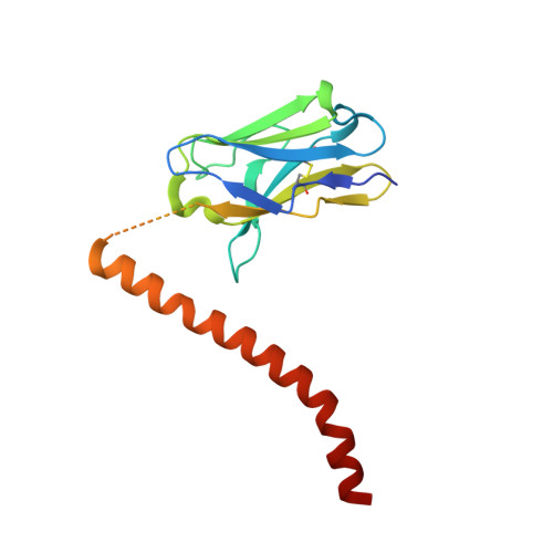
    Antibody fragments are frequently used as a "crystallization chaperone" to aid structural analysis of complex macromolecules that are otherwise crystallization resistant, but conventional fragment formats have not been designed for this particular application. By fusing an anti-parallel coiled-coil structure derived from the SARAH domain of human Mst1 kinase to the variable region of an antibody, we succeeded in creating a novel chimeric antibody fragment of ∼37 kDa, termed "Fv-clasp," which exhibits excellent crystallization compatibility while maintaining the binding ability of the original IgG molecule. The "clasp" and the engineered disulfide bond at the bottom of the Fv suppressed the internal mobility of the fragment and shielded hydrophobic residues, likely contributing to the high heat stability and the crystallizability of the Fv-clasp. Finally, Fv-clasp antibodies showed superior "chaperoning" activity over conventional Fab fragments, and facilitated the structure determination of an ectodomain fragment of integrin α6β1.


  • Organizational Affiliation

    Institute for Protein Research, Osaka University, Suita, Osaka 565-0871, Japan.


Macromolecules
Find similar proteins by:  (by identity cutoff)  |  3D Structure
Entity ID: 1
MoleculeChains Sequence LengthOrganismDetailsImage
VH(S112C)-SARAH chimera,VH(S112C)-SARAH chimera
A, D
172RattusHomo sapiens
This entity is chimeric
Mutation(s): 0 
Entity Groups  
Sequence Clusters30% Identity50% Identity70% Identity90% Identity95% Identity100% Identity
Sequence Annotations
Expand
  • Reference Sequence
Find similar proteins by:  (by identity cutoff)  |  3D Structure
Entity ID: 2
MoleculeChains Sequence LengthOrganismDetailsImage
VL-SARAH(S37C) chimera,VL-SARAH(S37C) chimera
B, E
165RattusHomo sapiens
This entity is chimeric
Mutation(s): 0 
Entity Groups  
Sequence Clusters30% Identity50% Identity70% Identity90% Identity95% Identity100% Identity
Sequence Annotations
Expand
  • Reference Sequence

Find similar proteins by:  Sequence   |   3D Structure  

Entity ID: 3
MoleculeChains Sequence LengthOrganismDetailsImage
PA14 peptide
C, F
14Homo sapiensMutation(s): 0 
UniProt & NIH Common Fund Data Resources
Find proteins for Q86YL7 (Homo sapiens)
Explore Q86YL7 
Go to UniProtKB:  Q86YL7
PHAROS:  Q86YL7
GTEx:  ENSG00000162493 
Entity Groups  
Sequence Clusters30% Identity50% Identity70% Identity90% Identity95% Identity100% Identity
UniProt GroupQ86YL7
Sequence Annotations
Expand
  • Reference Sequence
Experimental Data & Validation

Experimental Data

  • Method: X-RAY DIFFRACTION
  • Resolution: 2.14 Å
  • R-Value Free: 0.277 
  • R-Value Work: 0.228 
  • R-Value Observed: 0.231 
  • Space Group: C 1 2 1
Unit Cell:
Length ( Å )Angle ( ˚ )
a = 145.617α = 90
b = 50.122β = 110.46
c = 121.868γ = 90
Software Package:
Software NamePurpose
PHENIXrefinement
HKL-2000data reduction
HKL-2000data scaling
PHASERphasing

Structure Validation

View Full Validation Report


Entry History 

Deposition Data

Revision History  (Full details and data files)

  • Version 1.0: 2017-10-11
    Type: Initial release
  • Version 1.1: 2017-10-18
    Changes: Database references
  • Version 1.2: 2023-11-22
    Changes: Data collection, Database references, Derived calculations, Refinement description