5OBH

Crystal structure of glycine binding protein in complex with bicuculline


Experimental Data Snapshot

  • Method: X-RAY DIFFRACTION
  • Resolution: 2.40 Å
  • R-Value Free: 0.249 
  • R-Value Work: 0.230 
  • R-Value Observed: 0.231 

wwPDB Validation   3D Report Full Report


Ligand Structure Quality Assessment 


This is version 2.2 of the entry. See complete history


Literature

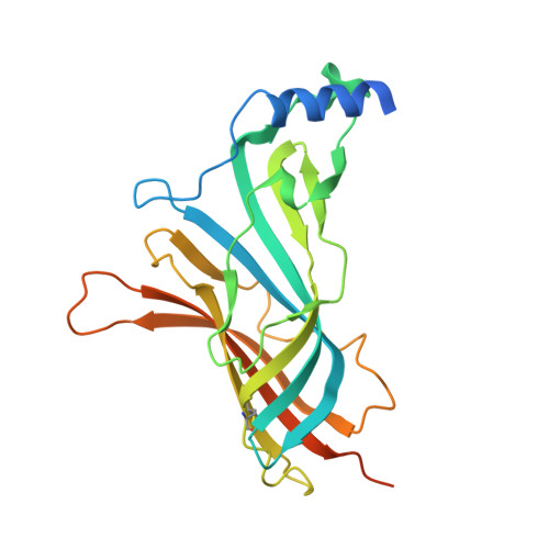
A Structural Rationale for N-Methylbicuculline Acting as a Promiscuous Competitive Antagonist of Inhibitory Pentameric Ligand-Gated Ion Channels.

Jones, M.J.Dawson, A.Hales, T.G.Hunter, W.N.

(2020) Chembiochem 21: 1526-1533

  • DOI: https://doi.org/10.1002/cbic.201900680
  • Primary Citation of Related Structures:  
    5OBH

  • PubMed Abstract: 

    Bicuculline, a valued chemical tool in neurosciences research, is a competitive antagonist of specific GABA A receptors and affects other pentameric ligand-gated ion channels including the glycine, nicotinic acetylcholine and 5-hydroxytryptamine type 3 receptors. We used a fluorescence-quenching assay and isothermal titration calorimetry to record low-micromolar dissociation constants for N-methylbicuculline interacting with acetylcholine-binding protein and an engineered version called glycine-binding protein (GBP), which provides a surrogate for the heteromeric interface of the extracellular domain of the glycine receptor (GlyR). The 2.4 Å resolution crystal structure of the GBP:N-methylbicuculline complex, sequence and structural alignments reveal similarities and differences between GlyR and the GABA A receptor-bicuculline interactions. N-methylbicuculline displays a similar conformation in different structures, but adopts distinct orientations enforced by interactions and steric blocks with key residues and plasticity in the binding sites. These features explain the promiscuous activity of bicuculline against the principal inhibitory pentameric ligand-gated ion channels in the CNS.


  • Organizational Affiliation

    Division of Biological Chemistry and Drug Discovery School of Life Sciences, University of Dundee, Dow St, Dundee, DD1 5EH, UK.


Macromolecules
Find similar proteins by:  (by identity cutoff)  |  3D Structure
Entity ID: 1
MoleculeChains Sequence LengthOrganismDetailsImage
Soluble acetylcholine receptor
A, B, C, D, E
249Aplysia californicaMutation(s): 9 
UniProt
Find proteins for Q8WSF8 (Aplysia californica)
Explore Q8WSF8 
Go to UniProtKB:  Q8WSF8
Entity Groups  
Sequence Clusters30% Identity50% Identity70% Identity90% Identity95% Identity100% Identity
UniProt GroupQ8WSF8
Sequence Annotations
Expand
  • Reference Sequence
Oligosaccharides

Help

Entity ID: 2
MoleculeChains Length2D Diagram Glycosylation3D Interactions
2-acetamido-2-deoxy-beta-D-glucopyranose-(1-4)-2-acetamido-2-deoxy-beta-D-glucopyranose-(1-4)-2-acetamido-2-deoxy-beta-D-glucopyranose
F
3N-Glycosylation
Glycosylation Resources
GlyTouCan:  G47362BJ
GlyCosmos:  G47362BJ
GlyGen:  G47362BJ
Small Molecules
Ligands 3 Unique
IDChains Name / Formula / InChI Key2D Diagram3D Interactions
J94 (Subject of Investigation/LOI)
Query on J94

Download Ideal Coordinates CCD File 
G [auth A],
I [auth B],
K [auth C],
M [auth D],
P [auth E]
(5S)-6,6-dimethyl-5-[(6R)-8-oxo-6,8-dihydrofuro[3,4-e][1,3]benzodioxol-6-yl]-5,6,7,8-tetrahydro[1,3]dioxolo[4,5-g]isoquinolin-6-ium
C21 H20 N O6
WDIQXKYUSINZME-RBUKOAKNSA-N
NAG
Query on NAG

Download Ideal Coordinates CCD File 
N [auth D],
Q [auth E]
2-acetamido-2-deoxy-beta-D-glucopyranose
C8 H15 N O6
OVRNDRQMDRJTHS-FMDGEEDCSA-N
CL
Query on CL

Download Ideal Coordinates CCD File 
H [auth A],
J [auth B],
L [auth C],
O [auth D],
R [auth E]
CHLORIDE ION
Cl
VEXZGXHMUGYJMC-UHFFFAOYSA-M
Biologically Interesting Molecules (External Reference) 1 Unique
Experimental Data & Validation

Experimental Data

  • Method: X-RAY DIFFRACTION
  • Resolution: 2.40 Å
  • R-Value Free: 0.249 
  • R-Value Work: 0.230 
  • R-Value Observed: 0.231 
  • Space Group: P 21 21 21
Unit Cell:
Length ( Å )Angle ( ˚ )
a = 71.243α = 90
b = 132.236β = 90
c = 132.631γ = 90
Software Package:
Software NamePurpose
REFMACrefinement
XDSdata reduction
Aimlessdata scaling
PHASERphasing

Structure Validation

View Full Validation Report


Ligand Structure Quality Assessment 


Entry History 

Deposition Data

Revision History  (Full details and data files)

  • Version 1.0: 2018-08-01
    Type: Initial release
  • Version 2.0: 2020-07-29
    Type: Remediation
    Reason: Carbohydrate remediation
    Changes: Atomic model, Data collection, Derived calculations, Structure summary
  • Version 2.1: 2021-06-30
    Changes: Database references, Structure summary
  • Version 2.2: 2024-01-17
    Changes: Data collection, Database references, Refinement description