


THE CRYSTAL STRUCTURE OF IGE FC MUTANT - P333C
Dhaliwal, B., Pang, M.O.Y., Keeble, A.H., Taylor, A.I., James, L.K., Gould, H.J., McDonnell, J.M., Beavil, A.J., Sutton, B.J.To be published.
Experimental Data Snapshot
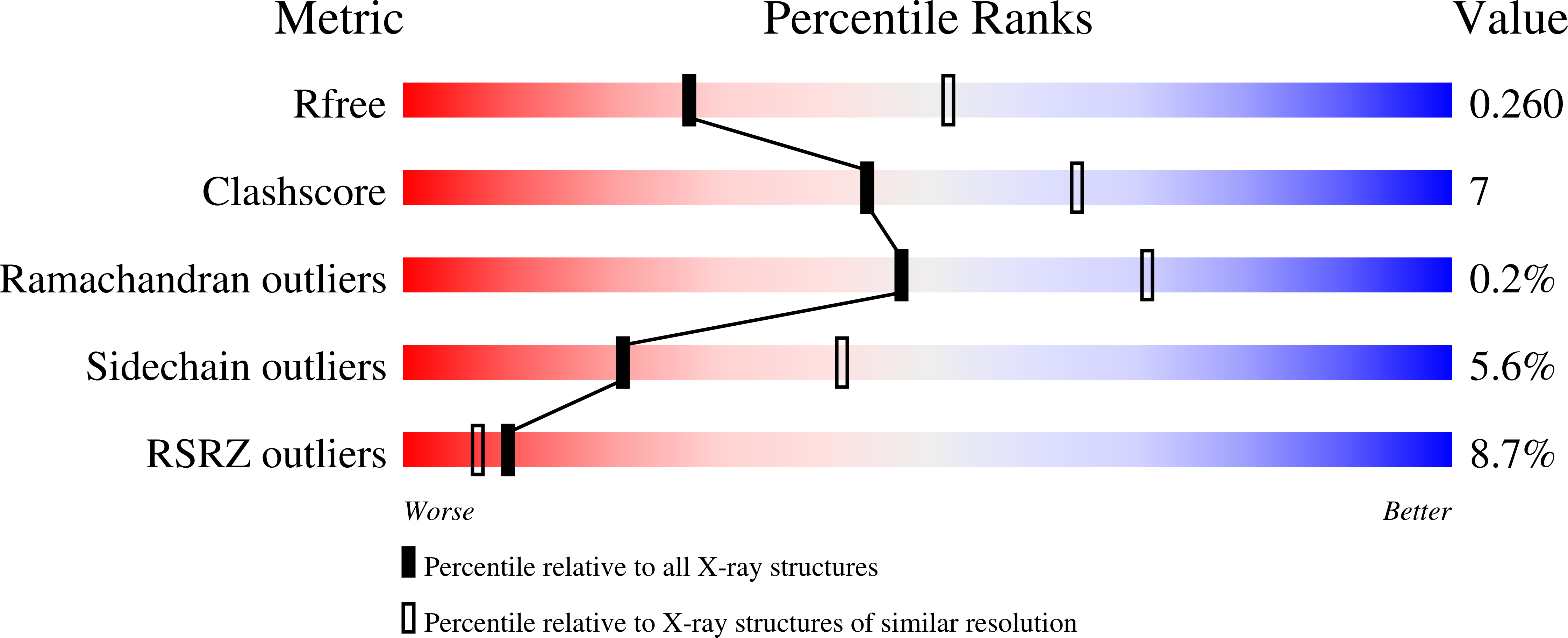
wwPDB Validation 3D Report Full Report
Entity ID: 1 | |||||
|---|---|---|---|---|---|
| Molecule | Chains | Sequence Length | Organism | Details | Image |
| Ig epsilon chain C region | 319 | Homo sapiens | Mutation(s): 1 Gene Names: IGHE | 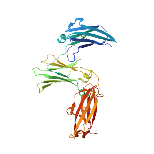 | |
UniProt & NIH Common Fund Data Resources | |||||
Find proteins for P01854 (Homo sapiens) Explore P01854 Go to UniProtKB: P01854 | |||||
PHAROS: P01854 GTEx: ENSG00000211891 | |||||
Entity Groups | |||||
| Sequence Clusters | 30% Identity50% Identity70% Identity90% Identity95% Identity100% Identity | ||||
| UniProt Group | P01854 | ||||
Sequence AnnotationsExpand | |||||
| |||||
Entity ID: 2 | |||||
|---|---|---|---|---|---|
| Molecule | Chains | Sequence Length | Organism | Details | Image |
| Ig epsilon chain C region | 320 | Homo sapiens | Mutation(s): 1 Gene Names: IGHE | 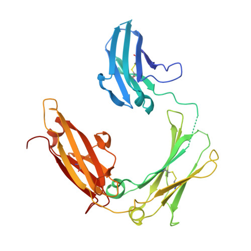 | |
UniProt & NIH Common Fund Data Resources | |||||
Find proteins for P01854 (Homo sapiens) Explore P01854 Go to UniProtKB: P01854 | |||||
PHAROS: P01854 GTEx: ENSG00000211891 | |||||
Entity Groups | |||||
| Sequence Clusters | 30% Identity50% Identity70% Identity90% Identity95% Identity100% Identity | ||||
| UniProt Group | P01854 | ||||
Sequence AnnotationsExpand | |||||
| |||||
Entity ID: 3 | |||||
|---|---|---|---|---|---|
| Molecule | Chains | Length | 2D Diagram | Glycosylation | 3D Interactions |
| alpha-D-mannopyranose-(1-3)-alpha-D-mannopyranose-(1-6)-beta-D-mannopyranose-(1-4)-2-acetamido-2-deoxy-beta-D-glucopyranose-(1-4)-2-acetamido-2-deoxy-beta-D-glucopyranose | C | 5 |  | N-Glycosylation | |
Glycosylation Resources | |||||
GlyTouCan: G10756ZZ GlyCosmos: G10756ZZ GlyGen: G10756ZZ | |||||
Entity ID: 4 | |||||
|---|---|---|---|---|---|
| Molecule | Chains | Length | 2D Diagram | Glycosylation | 3D Interactions |
| alpha-D-mannopyranose-(1-3)-[alpha-D-mannopyranose-(1-4)]alpha-D-mannopyranose-(1-6)-[alpha-D-mannopyranose-(1-3)]beta-D-mannopyranose-(1-4)-2-acetamido-2-deoxy-beta-D-glucopyranose-(1-4)-2-acetamido-2-deoxy-beta-D-glucopyranose | D | 7 |  | N-Glycosylation | |
Glycosylation Resources | |||||
GlyTouCan: G76464YC GlyCosmos: G76464YC GlyGen: G76464YC | |||||
| Ligands 3 Unique | |||||
|---|---|---|---|---|---|
| ID | Chains | Name / Formula / InChI Key | 2D Diagram | 3D Interactions | |
| PG4 Query on PG4 | H [auth A], I [auth A] | TETRAETHYLENE GLYCOL C8 H18 O5 UWHCKJMYHZGTIT-UHFFFAOYSA-N |  | ||
| SO4 Query on SO4 | E [auth A], F [auth A], G [auth A], K [auth B] | SULFATE ION O4 S QAOWNCQODCNURD-UHFFFAOYSA-L |  | ||
| GOL Query on GOL | J [auth B] | GLYCEROL C3 H8 O3 PEDCQBHIVMGVHV-UHFFFAOYSA-N |  | ||
| Length ( Å ) | Angle ( ˚ ) |
|---|---|
| a = 129.45 | α = 90 |
| b = 74.91 | β = 90 |
| c = 79.06 | γ = 90 |
| Software Name | Purpose |
|---|---|
| MOSFLM | data collection |
| Aimless | data scaling |
| PHASER | phasing |
| BUSTER-TNT | refinement |
| PDB_EXTRACT | data extraction |
| iMOSFLM | data reduction |
| PHASER | phasing |
| Funding Organization | Location | Grant Number |
|---|---|---|
| Medical Research Council (United Kingdom) | United Kingdom | -- |
| Wellcome Trust | United Kingdom | -- |
| Asthma UK | United Kingdom | -- |
RCSB PDB (citation) is hosted by
RCSB PDB is a member of the

