


THE CRYSTAL STRUCTURE OF IGE FC MUTANT - P333C
Dhaliwal, B., Pang, M.O.Y., Keeble, A.H., Taylor, A.I., James, L.K., Gould, H.J., McDonnell, J.M., Beavil, A.J., Sutton, B.J.To be published.
Experimental Data Snapshot
Starting Model: experimental
View more details
wwPDB Validation 3D Report Full Report

Entity ID: 1 | |||||
|---|---|---|---|---|---|
| Molecule | Chains | Sequence Length | Organism | Details | Image |
| Ig epsilon chain C region | 319 | Homo sapiens | Mutation(s): 1 Gene Names: IGHE | 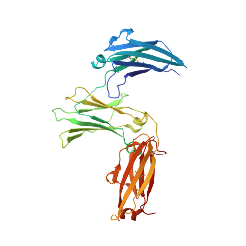 | |
UniProt & NIH Common Fund Data Resources | |||||
PHAROS: P01854 GTEx: ENSG00000211891 | |||||
Entity Groups | |||||
| Sequence Clusters | 30% Identity50% Identity70% Identity90% Identity95% Identity100% Identity | ||||
| UniProt Group | P01854 | ||||
Glycosylation | |||||
| Glycosylation Sites: 1 | Go to GlyGen: P01854-2 | ||||
Sequence AnnotationsExpand | |||||
Reference Sequence | |||||
Entity ID: 2 | |||||
|---|---|---|---|---|---|
| Molecule | Chains | Sequence Length | Organism | Details | Image |
| Ig epsilon chain C region | 320 | Homo sapiens | Mutation(s): 1 Gene Names: IGHE | 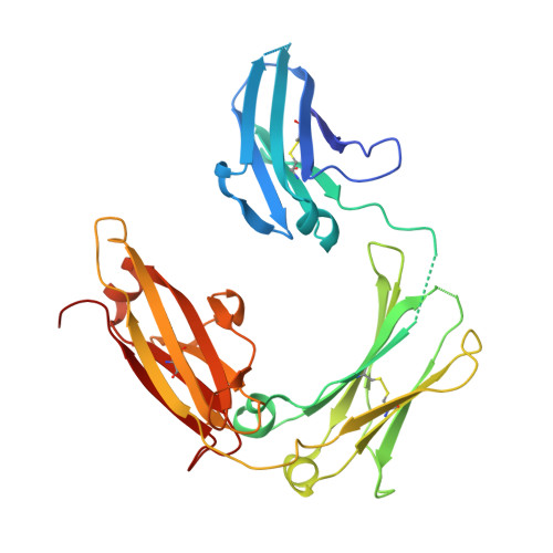 | |
UniProt & NIH Common Fund Data Resources | |||||
PHAROS: P01854 GTEx: ENSG00000211891 | |||||
Entity Groups | |||||
| Sequence Clusters | 30% Identity50% Identity70% Identity90% Identity95% Identity100% Identity | ||||
| UniProt Group | P01854 | ||||
Glycosylation | |||||
| Glycosylation Sites: 1 | Go to GlyGen: P01854-2 | ||||
Sequence AnnotationsExpand | |||||
Reference Sequence | |||||
Entity ID: 3 | |||||
|---|---|---|---|---|---|
| Molecule | Chains | Length | 2D Diagram | Glycosylation | D Interactions |
| alpha-D-mannopyranose-(1-3)-alpha-D-mannopyranose-(1-6)-beta-D-mannopyranose-(1-4)-2-acetamido-2-deoxy-beta-D-glucopyranose-(1-4)-2-acetamido-2-deoxy-beta-D-glucopyranose | C | 5 |  | N-Glycosylation | |
Glycosylation Resources | |||||
GlyTouCan: G10756ZZ GlyCosmos: G10756ZZ GlyGen: G10756ZZ | |||||
Entity ID: 4 | |||||
|---|---|---|---|---|---|
| Molecule | Chains | Length | 2D Diagram | Glycosylation | D Interactions |
| alpha-D-mannopyranose-(1-3)-[alpha-D-mannopyranose-(1-4)]alpha-D-mannopyranose-(1-6)-[alpha-D-mannopyranose-(1-3)]beta-D-mannopyranose-(1-4)-2-acetamido-2-deoxy-beta-D-glucopyranose-(1-4)-2-acetamido-2-deoxy-beta-D-glucopyranose | D | 7 |  | N-Glycosylation | |
Glycosylation Resources | |||||
GlyTouCan: G76464YC GlyCosmos: G76464YC GlyGen: G76464YC | |||||
| Ligands 3 Unique | |||||
|---|---|---|---|---|---|
| ID | Chains | Name / Formula / InChI Key | 2D Diagram | 3D Interactions | |
| PG4 Download:Ideal Coordinates CCD File | H [auth A], I [auth A] | TETRAETHYLENE GLYCOL C8 H18 O5 UWHCKJMYHZGTIT-UHFFFAOYSA-N |  | ||
| SO4 Download:Ideal Coordinates CCD File | E [auth A], F [auth A], G [auth A], K [auth B] | SULFATE ION O4 S QAOWNCQODCNURD-UHFFFAOYSA-L |  | ||
| GOL Download:Ideal Coordinates CCD File | J [auth B] | GLYCEROL C3 H8 O3 PEDCQBHIVMGVHV-UHFFFAOYSA-N |  | ||
| Length ( Å ) | Angle ( ˚ ) |
|---|---|
| a = 129.45 | α = 90 |
| b = 74.91 | β = 90 |
| c = 79.06 | γ = 90 |
| Software Name | Purpose |
|---|---|
| MOSFLM | data collection |
| Aimless | data scaling |
| PHASER | phasing |
| BUSTER-TNT | refinement |
| PDB_EXTRACT | data extraction |
| iMOSFLM | data reduction |
| PHASER | phasing |
| Funding Organization | Location | Grant Number |
|---|---|---|
| Medical Research Council (United Kingdom) | United Kingdom | -- |
| Wellcome Trust | United Kingdom | -- |
| Asthma UK | United Kingdom | -- |


