5KN8

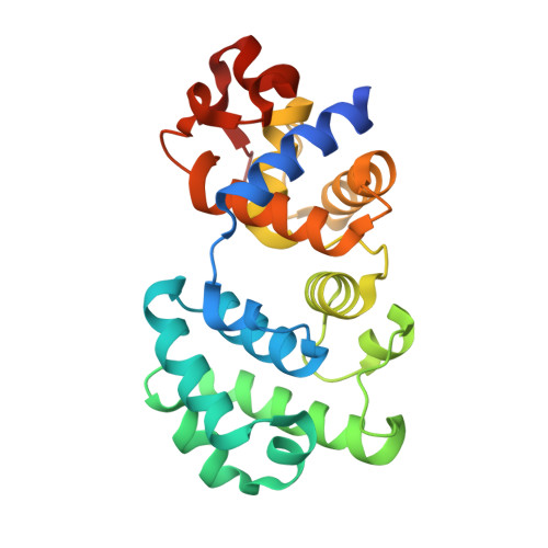
MutY N-terminal domain in complex with undamaged DNA


Experimental Data Snapshot

  • Method: X-RAY DIFFRACTION
  • Resolution: 1.81 Å
  • R-Value Free: 0.220 
  • R-Value Work: 0.167 
  • R-Value Observed: 0.169 

wwPDB Validation   3D Report Full Report


This is version 1.5 of the entry. See complete history


Literature

Structural Basis for the Lesion-scanning Mechanism of the MutY DNA Glycosylase.

Wang, L.Chakravarthy, S.Verdine, G.L.

(2017) J Biol Chem 292: 5007-5017

  • DOI: https://doi.org/10.1074/jbc.M116.757039
  • Primary Citation of Related Structures:  
    5KN8, 5KN9

  • PubMed Abstract: 

    The highly mutagenic A:8-oxoguanine (oxoG) base pair is generated mainly by misreplication of the C:oxoG base pair, the oxidation product of the C:G base pair. The A:oxoG base pair is particularly insidious because neither base in it carries faithful information to direct the repair of the other. The bacterial MutY (MUTYH in humans) adenine DNA glycosylase is able to initiate the repair of A:oxoG by selectively cleaving the A base from the A:oxoG base pair. The difference between faithful repair and wreaking mutagenic havoc on the genome lies in the accurate discrimination between two structurally similar base pairs: A:oxoG and A:T. Here we present two crystal structures of the MutY N-terminal domain in complex with either undamaged DNA or DNA containing an intrahelical lesion. These structures have captured for the first time a DNA glycosylase scanning the genome for a damaged base in the very first stage of lesion recognition and the base extrusion pathway. The mode of interaction observed here has suggested a common lesion-scanning mechanism across the entire helix-hairpin-helix superfamily to which MutY belongs. In addition, small angle X-ray scattering studies together with accompanying biochemical assays have suggested a possible role played by the C-terminal oxoG-recognition domain of MutY in lesion scanning.


  • Organizational Affiliation

    From the Departments of Chemistry and Chemical Biology.


Macromolecules

Find similar proteins by:  (by identity cutoff)  |  3D Structure
Entity ID: 1
MoleculeChains Sequence LengthOrganismDetailsImage
Adenine DNA glycosylase232Geobacillus stearothermophilusMutation(s): 2 
Gene Names: mutY
EC: 3.2.2
UniProt
Find proteins for P83847 (Geobacillus stearothermophilus)
Explore P83847 
Go to UniProtKB:  P83847
Entity Groups  
Sequence Clusters30% Identity50% Identity70% Identity90% Identity95% Identity100% Identity
UniProt GroupP83847
Sequence Annotations
Expand
  • Reference Sequence

Find similar nucleic acids by:  Sequence   |   3D Structure  

Entity ID: 2
MoleculeChains LengthOrganismImage
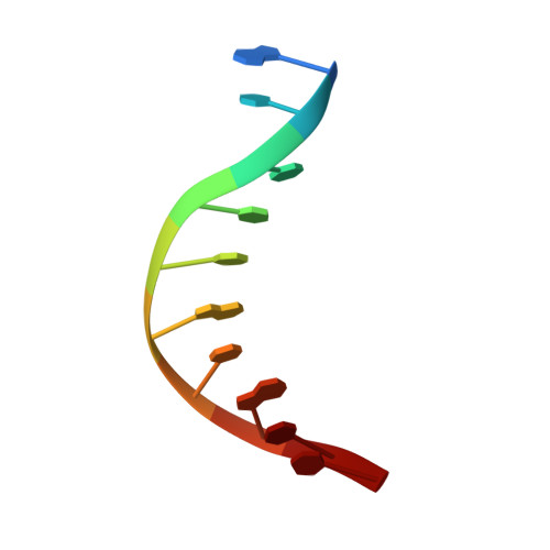
DNA (5'-D(*AP*TP*CP*CP*TP*GP*TP*GP*CP*T)-3')B [auth C]10Geobacillus stearothermophilus
Sequence Annotations
Expand
  • Reference Sequence

Find similar nucleic acids by:  Sequence   |   3D Structure  

Entity ID: 3
MoleculeChains LengthOrganismImage
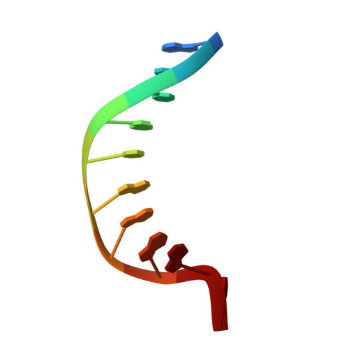
DNA (5'-D(*AP*GP*CP*AP*CP*AP*GP*GP*AP*T)-3')C [auth D]10Geobacillus stearothermophilus
Sequence Annotations
Expand
  • Reference Sequence
Small Molecules
Experimental Data & Validation

Experimental Data

  • Method: X-RAY DIFFRACTION
  • Resolution: 1.81 Å
  • R-Value Free: 0.220 
  • R-Value Work: 0.167 
  • R-Value Observed: 0.169 
  • Space Group: P 1 21 1
Unit Cell:
Length ( Å )Angle ( ˚ )
a = 49.2α = 90
b = 38.618β = 104
c = 83.796γ = 90
Software Package:
Software NamePurpose
PHENIXrefinement
XDSdata reduction
SCALAdata scaling
PHASERphasing

Structure Validation

View Full Validation Report


Entry History & Funding Information

Deposition Data


Funding OrganizationLocationGrant Number
National Institutes of Health/National Cancer Institute (NIH/NCI)United StatesCA100742

Revision History  (Full details and data files)

  • Version 1.0: 2017-02-08
    Type: Initial release
  • Version 1.1: 2017-02-22
    Changes: Database references
  • Version 1.2: 2017-04-05
    Changes: Database references
  • Version 1.3: 2017-09-27
    Changes: Author supporting evidence
  • Version 1.4: 2019-12-04
    Changes: Author supporting evidence
  • Version 1.5: 2023-09-27
    Changes: Data collection, Database references, Derived calculations, Refinement description