5J9A

Ambient temperature transition state structure of arginine kinase - crystal 11/Form II


Experimental Data Snapshot

  • Method: X-RAY DIFFRACTION
  • Resolution: 2.00 Å
  • R-Value Free: 0.179 
  • R-Value Work: 0.135 
  • R-Value Observed: 0.138 

wwPDB Validation   3D Report Full Report


Ligand Structure Quality Assessment 


This is version 1.5 of the entry. See complete history


Literature

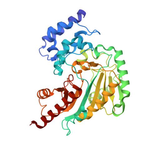
The Sampling of Conformational Dynamics in Ambient-Temperature Crystal Structures of Arginine Kinase.

Godsey, M.H.Davulcu, O.Nix, J.C.Skalicky, J.J.Bruschweiler, R.P.Chapman, M.S.

(2016) Structure 24: 1658-1667

  • DOI: https://doi.org/10.1016/j.str.2016.07.013
  • Primary Citation of Related Structures:  
    5J99, 5J9A

  • PubMed Abstract: 

    Arginine kinase provides a model for functional dynamics, studied through crystallography, enzymology, and nuclear magnetic resonance. Structures are now solved, at ambient temperature, for the transition state analog (TSA) complex. Analysis of quasi-rigid sub-domain displacements show that differences between the two TSA structures average about 5% of changes between substrate-free and TSA forms, and they are nearly co-linear. Small backbone hinge rotations map to sites that also flex on substrate binding. Anisotropic atomic displacement parameters (ADPs) are refined using rigid-body TLS constraints. Consistency between crystal forms shows that they reflect intrinsic molecular properties more than crystal lattice effects. In many regions, the favored directions of thermal/static displacement are appreciably correlated with movements on substrate binding. Correlation between ADPs and larger substrate-associated movements implies that the latter approximately follow paths of low-energy intrinsic motions.


  • Organizational Affiliation

    Department of Math/Science, Concordia University, Portland, OR 97211, USA.


Macromolecules
Find similar proteins by:  (by identity cutoff)  |  3D Structure
Entity ID: 1
MoleculeChains Sequence LengthOrganismDetailsImage
Arginine kinase357Limulus polyphemusMutation(s): 3 
EC: 2.7.3.3
UniProt
Find proteins for P51541 (Limulus polyphemus)
Explore P51541 
Go to UniProtKB:  P51541
Entity Groups  
Sequence Clusters30% Identity50% Identity70% Identity90% Identity95% Identity100% Identity
UniProt GroupP51541
Sequence Annotations
Expand
  • Reference Sequence
Experimental Data & Validation

Experimental Data

  • Method: X-RAY DIFFRACTION
  • Resolution: 2.00 Å
  • R-Value Free: 0.179 
  • R-Value Work: 0.135 
  • R-Value Observed: 0.138 
  • Space Group: P 21 21 21
Unit Cell:
Length ( Å )Angle ( ˚ )
a = 66.038α = 90
b = 72.047β = 90
c = 80.237γ = 90
Software Package:
Software NamePurpose
PHENIXrefinement
XDSdata reduction
XDSdata scaling
PHASERphasing

Structure Validation

View Full Validation Report


Ligand Structure Quality Assessment 


Entry History & Funding Information

Deposition Data


Funding OrganizationLocationGrant Number
National Institutes of Health/National Institute of General Medical Sciences (NIH/NIGMS)United StatesGM77643

Revision History  (Full details and data files)

  • Version 1.0: 2016-08-17
    Type: Initial release
  • Version 1.1: 2016-09-21
    Changes: Database references
  • Version 1.2: 2016-10-19
    Changes: Database references
  • Version 1.3: 2017-09-13
    Changes: Author supporting evidence, Database references, Derived calculations
  • Version 1.4: 2019-12-25
    Changes: Author supporting evidence
  • Version 1.5: 2023-09-27
    Changes: Data collection, Database references, Derived calculations, Refinement description